V2 Break Pattern
V2 break pattern
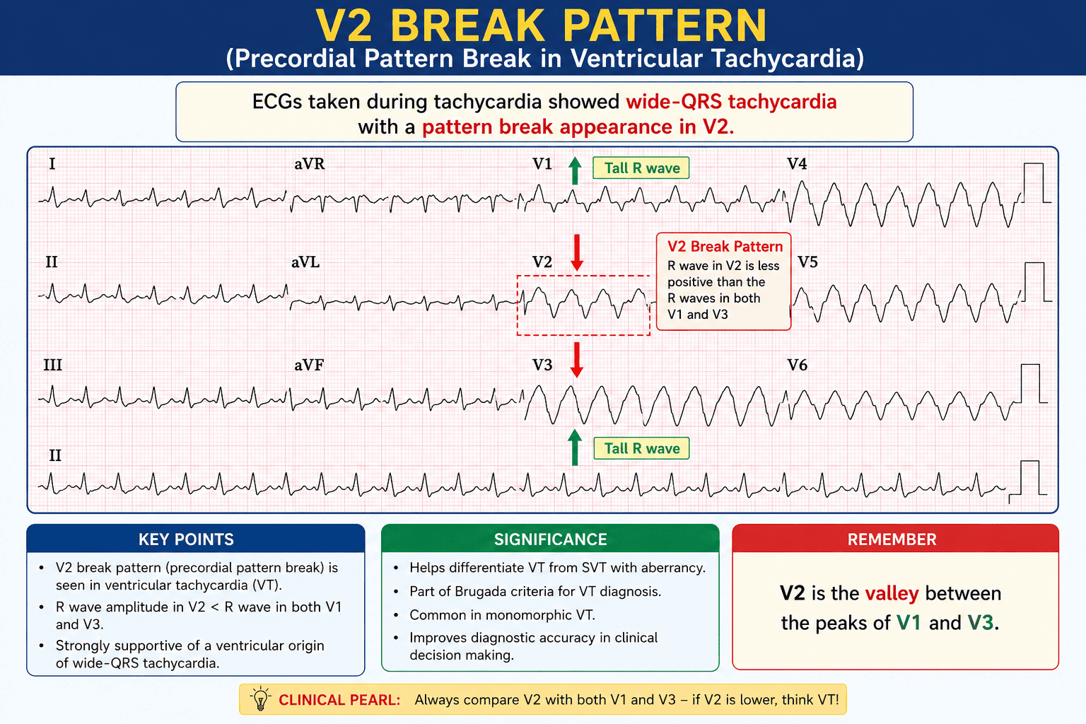
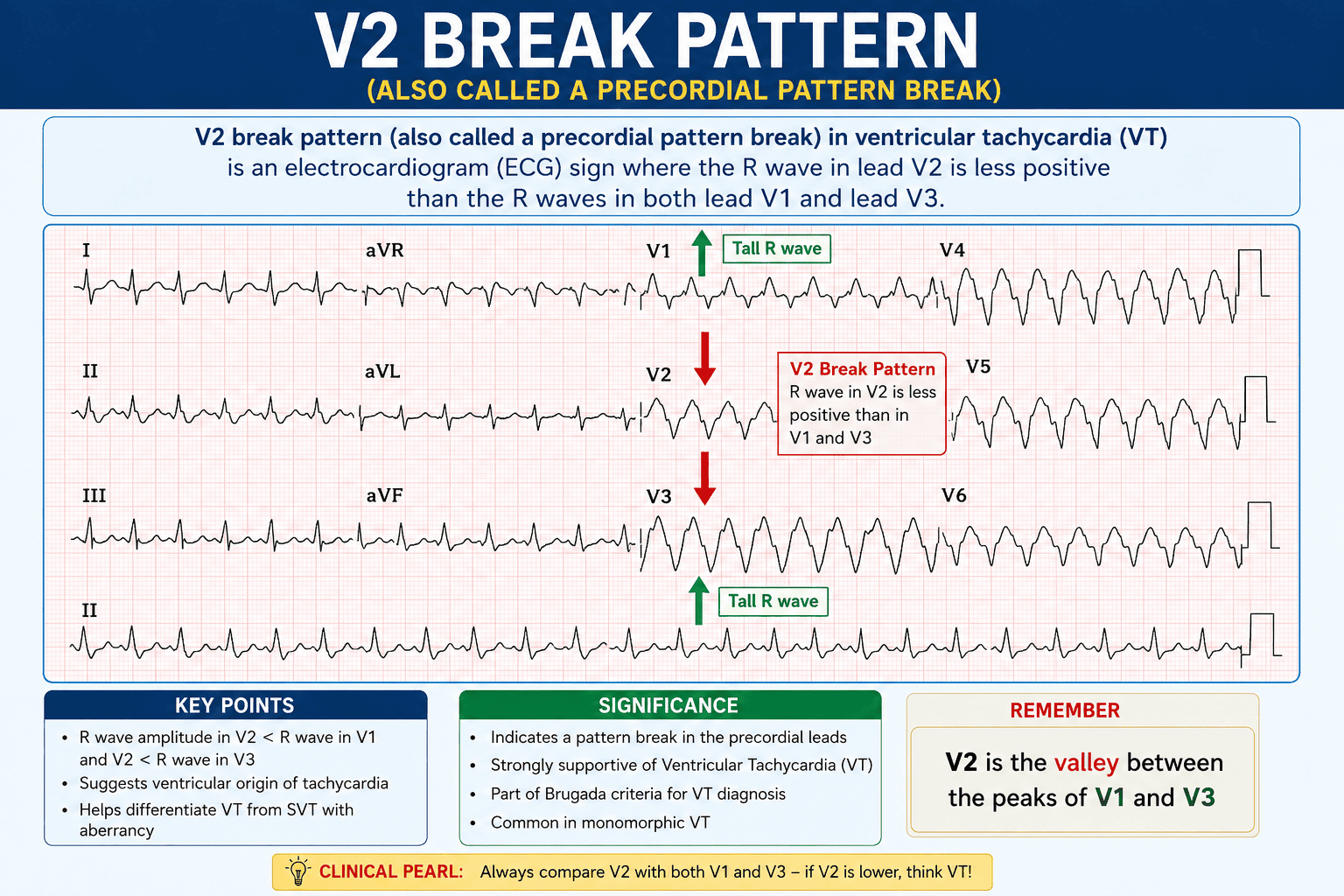
The V2 break pattern (also called a precordial pattern break) in ventricular tachycardia (VT) is an electrocardiogram (ECG) sign. V2 break pattern shows the R wave in lead V2 is less positive than the R waves in both lead V1 and lead V3. [1, 2]
Clinical Meaning
- Origin Site: It suggests that the ventricular arrhythmia comes from an epicardial location near the anterior interventricular sulcus or the left ventricular (LV) summit. This is close to the proximal left anterior descending (LAD) coronary artery. [1, 2]
- Electrical Vector: The depolarization vector runs in the opposite direction of unipolar lead V2. This cause an unexpected dip or QS pattern in that specific lead. [1]
Significance for Treatment
- Ablation Challenge: Arrhythmias with this sign are often harder to treat via catheter ablation. This is because the target site sits very close to major coronary blood vessels. [1]
A V2 pattern break (PBV2) on an electrocardiogram (ECG) is an unusual heart tracing sign showing an abrupt drop or loss of the R wave in lead V2 compared to leads V1 and V3. [1]
Key Features and Meaning
- The Sign: The R-wave amplitude in lead V2 is smaller or less positive than in both V1 and V3. [1, 2]
- Anatomy: It means the heart’s electrical signal starts near the anterior interventricular sulcus, which sits directly opposite lead V2 on the chest. [1, 2]
- Clinical Impact: Often seen in outflow tract ventricular arrhythmias (OT-VAs), making catheter ablation more difficult because the target area is very close to major blood vessels like the left anterior descending (LAD) coronary artery. [1, 2]
If you are researching this for a specific case, would you like to know more about catheter ablation success rates or the difference between standard and reverse pattern breaks?