Kayser-Fleischer Ring
🧠 Kayser–Fleischer Ring — Overview Definition:A Kayser–Fleischer ring is a brownish or golden-green discoloration encircling...
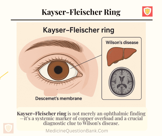
🧠 Kayser–Fleischer Ring — Overview Definition:A Kayser–Fleischer ring is a brownish or golden-green discoloration encircling...
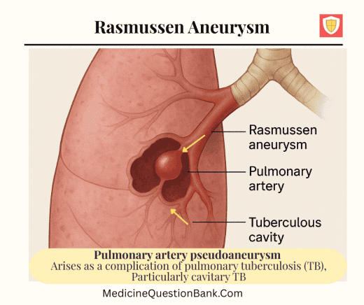
Rasmussen aneurysm 🩸 Rasmussen Aneurysm — Overview A Rasmussen aneurysm is a pulmonary artery pseudoaneurysm...
The “Triple Ripple” apical impulse (also known as Triple Apical Impulse or Triple Impulse) is...
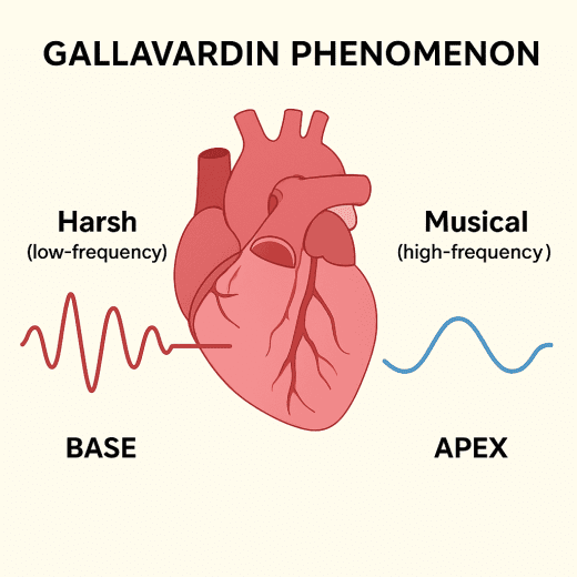
🫀 Gallavardin Phenomenon The Gallavardin phenomenon refers to the splitting of the murmur of aortic...
Ranson Score Calculator — Acute Pancreatitis .button-container { text-align: center; /* Centers the buttons horizontally...
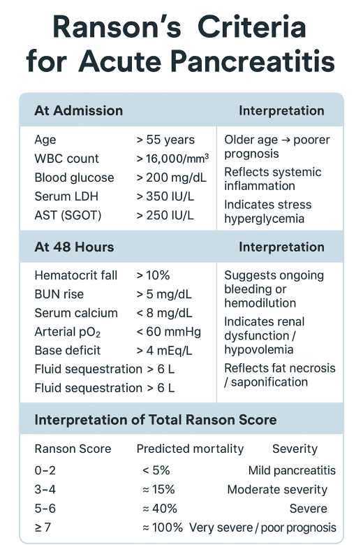
🩺 Ranson’s Criteria for Acute Pancreatitis Ranson’s Criteria for Acute Pancreatitis Ranson’s criteria help predict...
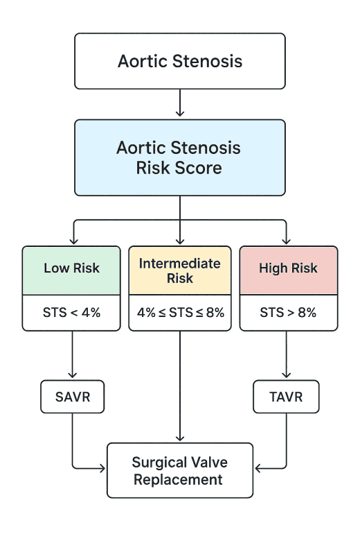
Aortic Stenosis Risk Score Calculator Aortic Stenosis Risk Score Interpretation Risk Category STS Predicted Mortality...
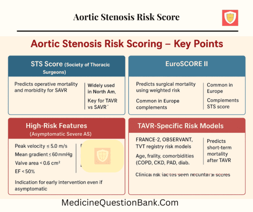
Aortic Stenosis Risk Score Main Risk Scores / Models Used in Aortic Stenosis 1. Surgical...
ADD-RS + D-dimer Calculator High-risk condition (Marfan, Ehlers-Danlos, bicuspid aortic valve, prior aortic surgery) High-risk...
Advanced USMLE-Style MCQs — ADD-RS & D-dimer Clinical vignettes with ADD-RS calculation, D-dimer interpretation, and...
Aortic Dissection Detection Risk Score (ADD-RS) and D-dimer: 🔹 Aortic Dissection Detection Risk Score (ADD-RS)...
Brockenbrough Phenomenon – USMLE-Style MCQ Q1. A 55-year-old man with a family history of sudden...
Brockenbrough phenomenon Brockenbrough–Braunwald–Morrow Sign — MCQs Q1. The Brockenbrough–Braunwald–Morrow sign is most classically associated with...
🫁 Kerley Lines Overview Kerley lines are thin linear pulmonary opacities seen on chest X-ray,...
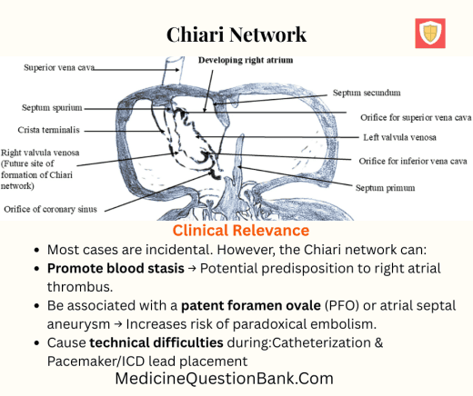
Chiari network Q1. The Chiari network is a remnant of which embryological structure? A. Eustachian...
Dock’s murmur Feature Description Definition A diastolic murmur heard in patients with severe coronary artery...
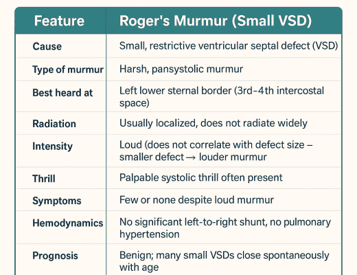
Roger’s murmur Feature Roger’s Murmur (Small VSD) Cause Small, restrictive ventricular septal defect (VSD) Type...
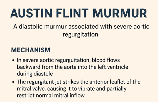
Austin Flint Murmur Austin Flint Murmur The Austin Flint murmur is a diastolic murmur associated...
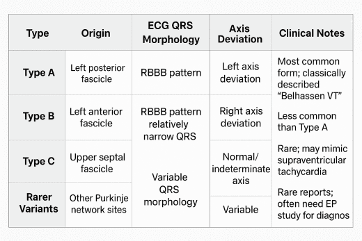
Belhassen Ventricular Tachycardia (VT) is a rare idiopathic form of ventricular tachycardia that typically arises...
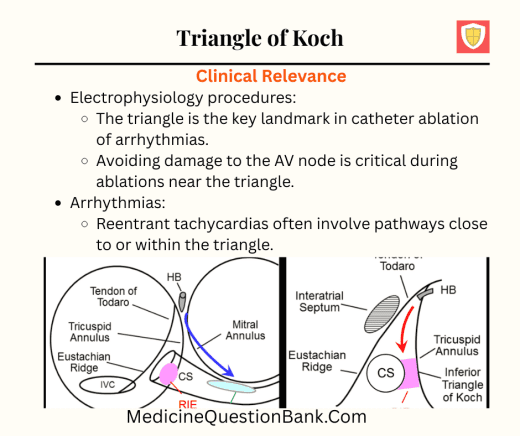
Triangle of Koch – Anatomy & Key Points Definition:The Triangle of Koch is an anatomical...
Tendon of Todaro – Anatomy & Details The tendon of Todaro is a small, collagenous band...
Cushing’s triad Cushing’s triad is a classic clinical sign of raised intracranial pressure (ICP), often...
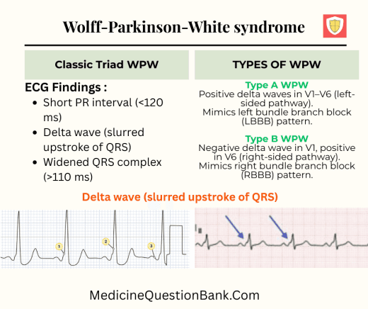
Types of Wolff–Parkinson–White (WPW) syndrome, we usually classify them based on pathway location, ECG pattern,...
WPW Syndrome Causes Symptoms WPW syndrome symptoms appear when a fast heartbeat occurs and vary...
Diagnostic criteria for left atrial abnormality and right atrial abnormality — based on ECG findings...
Aschoff body An Aschoff body (also called Aschoff nodule) is the pathognomonic lesion of rheumatic...
classification of coronary artery aneurysms (CAA) by size 📏 🔹 Size-Based Classification of CAA ✅...
Coronary artery aneurysms Coronary Artery Aneurysm (CAA) Definition Epidemiology Etiology Pathophysiology Clinical Features Diagnosis Complications...
Splinter Hemorrhage – Medical Overview Definition:Splinter hemorrhages are small, linear, red-to-brown streaks under the nails...
Low QRS Voltage in ECG 🔹 Definition Low QRS Voltage is defined when the amplitude...
1. The Gorlin formula is primarily used to calculate: Valve area in stenotic lesions Cardiac...
Carabello sign Carabello Sign – Clinical Cardiology The Carabello sign is a classic bedside clinical...
25-year-old woman on a vegetarian diet presented with lancinating pains mainly affecting the lower extremities. ...
What is the diagnosis? Scientific Figure on ResearchGate. Available from: https://www.researchgate.net/figure ‘Mercedes-Benz’ sign What is...
What is the diagnosis of the Image? Scientific Figure on Research Gate. A. Sunflower Cataract...
What is the diagnosis? What is the diagnosis of Image? A. Epidemic keratoconjunctivitis B. Vernal...
Where is the most probable site of the lesion if the following visual field defect...
A. Hensen’s cells B. Oxyphil cell C. Mott cell D. Club cell Mott cells Mott...
What is the diagnosis of the cell shown in Image? A. M-Cell B. Cart Wheel...
What is the diagnosis of the Marked structure in IMAGE? User:Plainpaper, CC BY-SA 3.0 https://creativecommons.org/licenses/by-sa/3.0,...
Forchheimer spots Forchheimer spots are ——- for rubella A. Specific B. Non specific C. Pathognomonic...
Presentation 61 year female presented with nonspecific complaints such as fatigue and dyspnea on exertion...
What is the diagnosis ? History 7 year male child presented to emergency department with...
Mickey Mouse sign Mickey Mouse sign is described on ultrasound image at the groin in...

FREE Updates, MCQs & Questions For Doctors & Medical Students