Image Question-35
Mickey Mouse sign
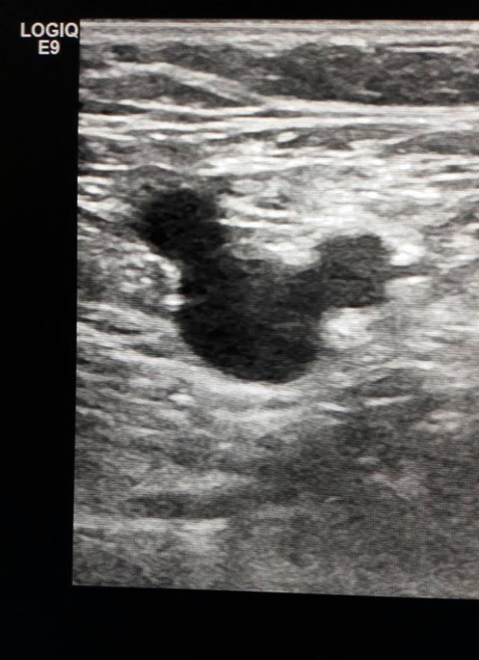
Mickey Mouse sign is described on ultrasound image at the groin in presence of dilated ———–
A. accessory saphenous vein
B. common femoral vein
C. great saphenous vein
D. common femoral artery
The presence of a Mickey Mouse sign has been a great diagnostic clue to check accessory saphenous vein insufficiency.
“Mickey Mouse View”
“Mickey Mouse View” at the saphenofemoral junction in the groin: the common femoral vein (CFV) represents the head of Mickey Mouse while the great saphenous vein (GSV) and the femoral artery (CFA) represent the ears
Mickey Mouse sign
| Mickey Mouse sign | |||
| 1 | Midbrain of progressive supranuclear palsy | MRI brain demonstrate | ‘Humming bird sign’, ‘Mickey mouse sign’, and ‘Morning glory sign’. |
| 2 | Polyostotic Paget’s disease | bone scintigraphy with technetium-99m | Increased scintigraphic uptake in the body and spine of the vertebrae resembles the head of Mickey Mouse |
| 3 | Portal triad | biliary ultrasound scans | portal vein comprising Mickey’s head and the common bile duct and hepatic artery his right and left ears, respectively |
| 4 | Pelvic Mickey Mouse sign | bilateral inguinal vesical hernia | on transverse axial imaging |
| 5 | Dysmorphic Mickey Mouse RBCs | presence of these dysmorphic cells in urine | indicate intraglomerular haemorrhage |
| 6 | Ureteropelvic junction obstruction | USG |


