Image Question-31
What is the Diagnosis of Image?
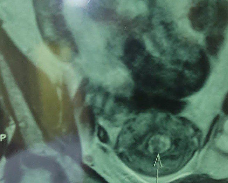
What is the Diagnosis of MRI Image?
A. Endometriosis
B. Carcinoma cervix
C. Adenomyoma of uterus
D. Tumor of Urinary bladder
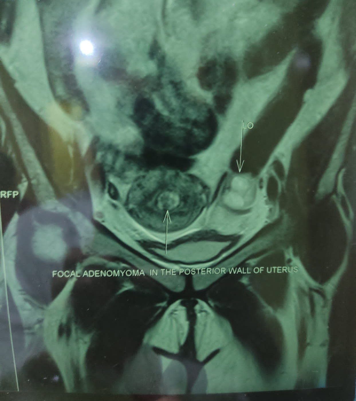
Adenomyoma – lesion is characteristically a well-circumscribed, gray or white mass that may contain multiple mucinous cysts.
Histologically it is composed of irregularly shaped glands, some of which may exhibit papillary infoldings, and a leaflike architecture surrounded by smaller rounded glands, imparting a lobular appearance.
How to distinguish Adenomyoma from a uterine fibroid?
Adenomyoma is a focal region of adenomyosis resulting in a mass, which is difficult to distinguish from a uterine fibroid.
- Adenomyoma – mass is poorly defined and blends with the surrounding myometrium.
- Uterine fibroids- have a pseudocapsule of compressed myometrial tissue surrounding them.


