Pulmonary Embolism (PE): 30 High-Yield Clinical, ECG, and Echocardiography Points
Pulmonary Embolism Clinical ECG and Echocardiography
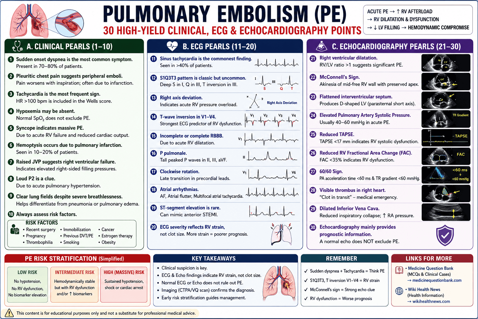
Pulmonary Embolism (PE): 30 High-Yield Clinical, ECG, and Echocardiography Points
A. Clinical Pearls (1–10)
1. Sudden onset dyspnea is the most common symptom.
- Present in approximately 70–80% of patients.
- Severity depends on clot burden and cardiopulmonary reserve.
2. Pleuritic chest pain suggests peripheral emboli.
- Pain worsens with inspiration.
- Often associated with pulmonary infarction.
3. Tachycardia is the most frequent physical sign.
- Heart rate >100 bpm is included in the Wells score.
4. Hypoxemia may be absent.
- A normal oxygen saturation does not exclude PE.
- Young patients may maintain normal oxygenation despite significant emboli.
5. Syncope indicates massive PE.
- Results from acute RV failure and reduced cardiac output.
- Associated with increased mortality.
6. Hemoptysis occurs due to pulmonary infarction.
- Seen in approximately 10–20% of patients.
- Usually accompanied by pleuritic pain.
7. Raised JVP suggests right ventricular failure.
- Indicates elevated right-sided filling pressures.
8. Loud P2 is a clue.
- Due to acute pulmonary hypertension.
9. Clear lung fields despite severe breathlessness.
- Helps differentiate PE from pneumonia or pulmonary edema.
10. Always assess risk factors.
- Recent surgery
- Immobilization
- Cancer
- Pregnancy
- Previous DVT/PE
- Estrogen therapy
- Thrombophilia
B. ECG Pearls (11–20)
11. Sinus tachycardia is the commonest ECG finding.
- Seen in >40% of patients.
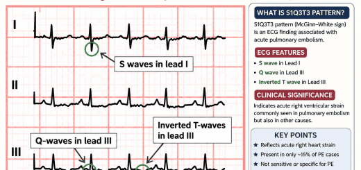
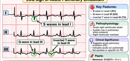
12. S1Q3T3 pattern is classic but uncommon.
- Deep S wave in Lead I
- Q wave in Lead III
- T-wave inversion in Lead III
- Suggests acute RV strain.
13. Right axis deviation
- Indicates acute RV pressure overload.
14. T-wave inversion in V1–V4
- One of the strongest ECG predictors of RV dysfunction.
- Correlates with worse prognosis.
15. Incomplete or complete Right Bundle Branch Block
- Due to acute RV dilatation.
16. P pulmonale
- Tall peaked P waves in II, III, aVF.
- Indicates right atrial enlargement.
17. Clockwise rotation
- Late transition in precordial leads.
18. Atrial arrhythmias
- Atrial fibrillation
- Atrial flutter
- Multifocal atrial tachycardia
- May occur in severe PE.
19. ST-segment elevation is rare.
- Can mimic anterior STEMI due to severe RV strain.
20. ECG severity reflects RV strain rather than clot size.
- More RV strain = poorer prognosis.
C. Echocardiography Pearls (21–30)
21. Right ventricular dilatation
- RV/LV ratio >1 suggests significant PE.
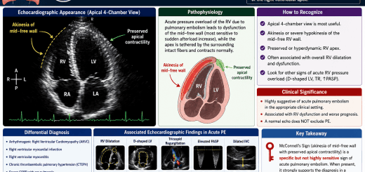
22. McConnell’s Sign
- Akinesia of mid-free RV wall
- Preserved RV apex
- Highly suggestive of acute PE.
23. Flattened interventricular septum
- Produces D-shaped LV.
- Indicates RV pressure overload.
24. Elevated Pulmonary Artery Systolic Pressure
- Usually 40–60 mmHg in acute PE.
- Very high pressures suggest chronic pulmonary hypertension.
25. Reduced TAPSE
- TAPSE <17 mm indicates RV systolic dysfunction.
- Poor prognostic marker.
26. Reduced RV Fractional Area Change (FAC)
- FAC <35% indicates RV dysfunction.
27. 60/60 Sign
- Pulmonary acceleration time <60 ms
- TR gradient <60 mmHg
- Suggestive of acute PE.
28. Visible thrombus in right heart
- “Clot in transit.”
- Medical emergency with very high mortality.
29. Dilated Inferior Vena Cava
- Reduced inspiratory collapse.
- Reflects elevated RA pressure.
30. Echocardiography mainly provides prognostic information.
- A normal echocardiogram does not exclude PE.
- In unstable patients with shock, evidence of RV strain on echo supports immediate reperfusion therapy when PE is strongly suspected.
High-Yield One-Liners for Exams
| Finding | Clinical Importance |
|---|---|
| Sudden dyspnea | Most common symptom |
| Tachycardia | Most common sign |
| Syncope | Massive PE until proven otherwise |
| Hemoptysis | Pulmonary infarction |
| Clear chest | Common despite severe PE |
| S1Q3T3 | Classic RV strain pattern |
| Sinus tachycardia | Most common ECG finding |
| T inversion V1–V4 | Strong marker of RV strain |
| RBBB | Acute RV overload |
| Right axis deviation | RV pressure overload |
| McConnell sign | Suggestive of acute PE |
| RV/LV >1 | Significant RV dysfunction |
| D-shaped LV | RV pressure overload |
| TAPSE <17 mm | RV dysfunction |
| FAC <35% | RV systolic dysfunction |
| 60/60 sign | Acute PE clue |
| Clot in transit | Very high mortality |
| Dilated IVC | Elevated RA pressure |
| Normal ECG | Does not exclude PE |
| Normal Echo | Does not rule out PE |
Clinical Pearls
- Massive PE: Sustained hypotension, shock, or cardiac arrest.
- Intermediate-risk (submassive) PE: Hemodynamically stable but with RV dysfunction and/or elevated cardiac biomarkers.
- Low-risk PE: No hypotension, no RV dysfunction, and no biomarker elevation.
- CT Pulmonary Angiography (CTPA) is the imaging test of choice in hemodynamically stable patients.
- In hemodynamically unstable patients, bedside echocardiography demonstrating acute RV strain can guide urgent reperfusion therapy when immediate CTPA is not feasible.
SEO Meta Table – Pulmonary Embolism: 30 Clinical, ECG and Echocardiography Points
| SEO Element | Content |
|---|---|
| SEO Title | Pulmonary Embolism: 30 Clinical, ECG & Echocardiography Pearls for Students and Clinicians |
| Focus Keyword | Pulmonary Embolism Clinical ECG and Echocardiography |
| Secondary Keywords | Pulmonary embolism ECG findings, PE echocardiography, right ventricular strain, McConnell sign, S1Q3T3, pulmonary embolism diagnosis, acute pulmonary embolism, RV dysfunction in PE |
| SEO URL (Slug) | pulmonary-embolism-clinical-ecg-echo-points |
| Meta Description (155–160 characters) | Learn 30 high-yield clinical, ECG, and echocardiographic findings of pulmonary embolism, including RV strain, McConnell sign, S1Q3T3, and bedside diagnostic pearls. |
| Meta Keywords | Pulmonary embolism, PE ECG, Echocardiography PE, McConnell sign, RV strain, S1Q3T3, TAPSE, D-shaped LV, pulmonary hypertension, cardiology notes |
| Article Category | Cardiology |
| Content Type | Clinical Review |
| Reading Time | 6–8 minutes |
| Target Audience | MBBS Students, MD Medicine, DM Cardiology, NEET-PG, NEET-SS, Residents, Emergency Physicians |
| Schema Type | MedicalWebPage, Article, FAQ |
| Image Alt Text | Echocardiographic and ECG findings in acute pulmonary embolism |
| Image File Name | pulmonary-embolism-ecg-echo-clinical-findings.webp |
Internal Links
- Medicine Question Bank (Clinical MCQs & Exam Preparation): Medicine Question Bank
- Suggested anchor text:
- Pulmonary Embolism MCQs
- ECG Interpretation
- Echocardiography Basics
- Emergency Cardiology
- Right Ventricular Dysfunction
External Link
- General health education: Wiki Health News
- Pulmonary Embolism Awareness
- Heart and Lung Health
- Prevention of Deep Vein Thrombosis
- Healthy Lifestyle for Cardiovascular Disease
The sites above provide medical education and health information resources. (medicinequestionbank.com)