Triangle of Koch
Triangle of Koch – Anatomy & Key Points
Definition:
The Triangle of Koch is an anatomical area in the right atrium of the heart that is critically important in cardiac electrophysiology because it contains the atrioventricular (AV) node.
Boundaries
The triangle is roughly triangular in shape and is defined by three anatomical landmarks:
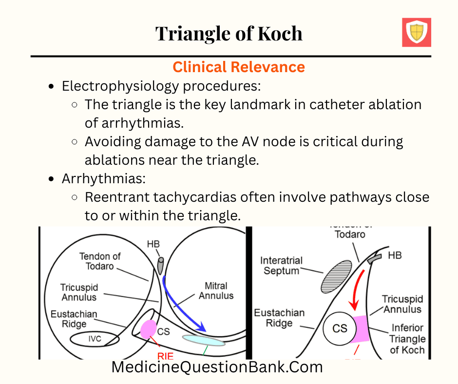
- Tendon of Todaro (superior border)
- Fibrous structure extending from the Eustachian valve to the central fibrous body.
- Important as a landmark for locating the AV node.
- Coronary sinus ostium (posterior-inferior border)
- The opening of the coronary sinus into the right atrium.
- Septal leaflet of the tricuspid valve (anterior-inferior border)
- The part of the tricuspid valve attached to the interventricular septum.
Contents
- AV node: The main conduction node between atria and ventricles.
- His bundle origin: The AV bundle arises near the apex of the triangle.
Clinical Relevance
- Electrophysiology procedures:
- The triangle is the key landmark in catheter ablation of arrhythmias.
- Avoiding damage to the AV node is critical during ablations near the triangle.
- Arrhythmias:
- Reentrant tachycardias often involve pathways close to or within the triangle.
Mnemonic
- “TAC” for Triangle of Koch:
- T = Tendon of Todaro
- A = AV node (apex)
- C = Coronary sinus ostium
Explanation: The Triangle of Koch is in the right atrium and contains the AV node.
Explanation: The tendon of Todaro forms the superior border.
Explanation: The coronary sinus ostium forms the posterior-inferior boundary.
Explanation: The septal leaflet of the tricuspid valve forms the anterior-inferior border.
Explanation: The AV node lies at the apex.
Explanation: Tendon of Todaro connects the Eustachian valve to the central fibrous body.
Explanation: Triangle of Koch contains the AV node, critical for conduction.
Explanation: The His bundle originates at the apex of the Triangle of Koch.
Explanation: Pulmonary valve is not part of the Triangle of Koch.
Explanation: AV node lies within the Triangle of Koch.
Explanation: Tendon of Todaro is used as an anatomical landmark to identify the AV node region.
Explanation: The septal leaflet of the tricuspid valve lies lateral to the triangle.
Explanation: The coronary sinus ostium forms the posterior-inferior border.
Explanation: The Tendon of Todaro is a fibrous structure, not muscular or nodal tissue.
Explanation: The AV node is located at the apex of the triangle.
Explanation: The anterior-inferior border is formed by the septal leaflet.
Explanation: The AV node lies at the apex of the triangle.
Explanation: Tendon of Todaro is used as a landmark to locate the AV node.
Explanation: Pulmonary valve is not part of the triangle.
Explanation: Tendon of Todaro is a fibrous landmark used in electrophysiology to locate the AV node.
Triangle of Koch, Koch's triangle, right atrium, atrioventricular node, AV node, tendon of Todaro, coronary sinus ostium, septal leaflet of tricuspid valve, atrial septum, membranous septum, AV nodal artery, His bundle, compact AV node, slow pathway, fast pathway, nodal extensions, posterior septal region, conduction system, arrhythmia substrate, ablation target


