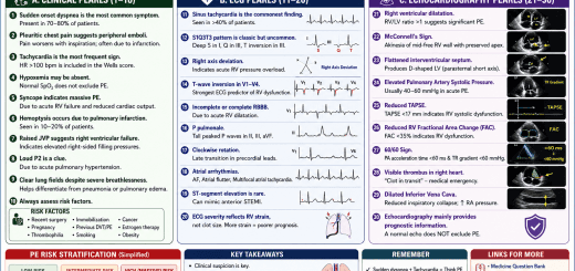
McConnell’s Sign Echocardiography Guide
McConnell’s Sign Echocardiography Guide
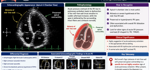
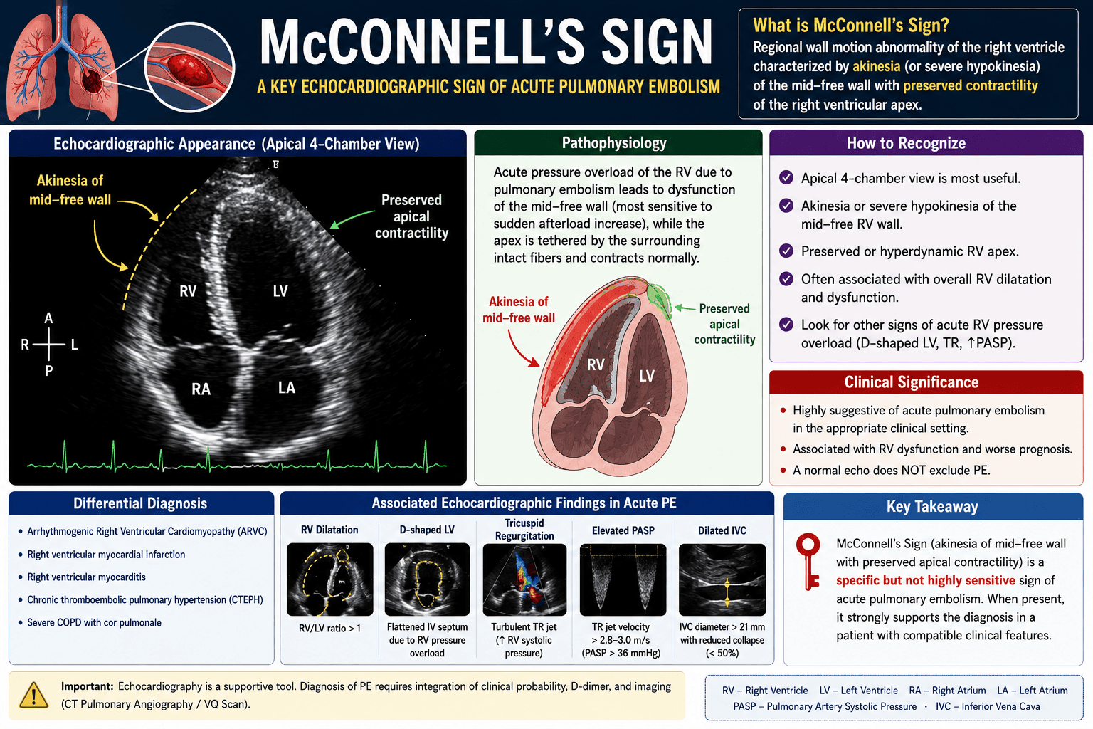
McConnell’s Sign is a distinctive echocardiographic finding characterized by akinesia or severe hypokinesia of the mid-free wall of the right ventricle (RV) with preserved or hyperdynamic contraction of the RV apex. It is an important marker of acute right ventricular pressure overload, most commonly associated with acute pulmonary embolism (PE).
Definition
McConnell’s sign is defined as:
- Akinesia (or severe hypokinesia) of the mid-free wall of the right ventricle
- Preserved or hyperdynamic motion of the RV apex
- Best visualized in the apical four-chamber (A4C) echocardiographic view
Pathophysiology
Acute pulmonary embolism causes a sudden increase in pulmonary vascular resistance, leading to:
- Acute RV pressure overload
- Right ventricular dilatation
- Reduced contraction of the mid-free wall
- Preserved apical contraction due to tethering from the left ventricle and relatively preserved apical myocardial perfusion
This unique regional wall-motion pattern produces McConnell’s sign.
How to Identify McConnell’s Sign
Echocardiographic View
- Apical four-chamber view (preferred)
Diagnostic Features
- Dilated right ventricle
- Mid-RV free-wall akinesia or severe hypokinesia
- Normal or hyperdynamic RV apical contraction
- Reduced global RV systolic function
Clinical Significance
McConnell’s sign is highly suggestive of acute pulmonary embolism in the appropriate clinical setting.
It indicates:
- Significant RV pressure overload
- Right ventricular dysfunction
- Increased risk of hemodynamic deterioration
- Higher short-term mortality in acute PE
Associated Echocardiographic Findings
Patients with acute PE often have additional findings:
- RV/LV basal diameter ratio >1.0
- D-shaped left ventricle due to septal flattening
- Dilated inferior vena cava with reduced inspiratory collapse
- Moderate tricuspid regurgitation
- Elevated pulmonary artery systolic pressure
- Reduced TAPSE (<17 mm)
- Reduced RV fractional area change (<35%)
- 60/60 sign (pulmonary acceleration time <60 ms with TR gradient <60 mmHg)
- Right-heart thrombus (“clot in transit”) in severe cases
Conditions That Can Mimic McConnell’s Sign
McConnell’s sign is not specific to pulmonary embolism alone and may occasionally be seen in:
- Right ventricular myocardial infarction
- Chronic thromboembolic pulmonary hypertension (CTEPH)
- Arrhythmogenic right ventricular cardiomyopathy (ARVC)
- Severe pulmonary hypertension
- Right ventricular myocarditis
Diagnostic Accuracy
| Parameter | Approximate Value |
|---|---|
| Sensitivity | 20–40% |
| Specificity | 85–95% |
| Positive Predictive Value | Higher when clinical suspicion of PE is high |
Key point: McConnell’s sign is more specific than sensitive. Its absence does not exclude pulmonary embolism.
Role in Acute Pulmonary Embolism
In patients with:
- Shock
- Persistent hypotension
- Cardiac arrest
- Severe hypoxemia
McConnell’s sign supports the diagnosis of high-risk (massive) pulmonary embolism, especially when immediate CT pulmonary angiography is not feasible. In this setting, bedside echocardiography can help guide urgent reperfusion decisions while integrating the overall clinical picture.
Limitations
- Not pathognomonic for pulmonary embolism
- Requires good image quality
- May be confused with RV infarction
- Should never be interpreted in isolation
- Confirmation with CT pulmonary angiography (or V/Q scan when appropriate) is recommended in hemodynamically stable patients
Key Examination Pearls
- Best view: Apical four-chamber
- Classic finding: Mid-RV free-wall akinesia with preserved RV apex
- Suggests: Acute pulmonary embolism with RV strain
- Associated findings: RV dilatation, D-shaped LV, elevated PASP, reduced TAPSE
- Normal echocardiogram does not exclude PE
- McConnell’s sign should always be interpreted alongside clinical assessment, ECG, biomarkers, and definitive imaging
| Content |
|---|
| McConnell’s Sign Echocardiography Guide: Diagnosis of Acute Pulmonary Embolism |
| McConnell’s Sign Echocardiography Guide |
| Learn how to identify McConnell’s sign on echocardiography, its pathophysiology, diagnostic value, limitations, and role in acute pulmonary embolism. |
| McConnell sign, RV strain, pulmonary embolism echocardiography, right ventricular dysfunction, acute PE echo, apical four-chamber view |
| McConnell’s sign on echocardiography showing mid-right ventricular free-wall akinesia with preserved apical contraction |
| https://medicinequestionbank.com/ (Pulmonary Embolism MCQs, Echocardiography Notes, Emergency Cardiology) |
| https://wikihealthnews.com/ (Pulmonary Embolism Awareness and Heart Health) |
1. McConnell’s sign is classically associated with?
McConnell’s sign is a classic echocardiographic finding in acute pulmonary embolism and consists of regional right ventricular dysfunction with preserved apical contraction.
2. McConnell’s sign is characterized by:
The hallmark of McConnell’s sign is akinesia or hypokinesia of the RV free wall while the RV apex remains normally contractile.
3. Which echocardiographic view best demonstrates McConnell’s sign?
The apical four-chamber view provides the best visualization of regional RV free wall motion abnormalities and preserved apical contraction.
4. The proposed mechanism behind preserved RV apical motion is:
The RV apex remains contractile because it is mechanically tethered to the normally contracting LV apex despite RV free wall dysfunction.
5. Which statement regarding McConnell’s sign is TRUE?
Although McConnell’s sign strongly suggests acute pulmonary embolism, it is not completely specific and can also be observed in right ventricular myocardial infarction and other causes of acute RV dysfunction.
6. Which chamber primarily demonstrates the abnormal wall motion in McConnell’s sign?
McConnell’s sign is a regional wall-motion abnormality of the right ventricle (RV), characterized by hypokinesia of the RV free wall with preserved apical contraction.
7. McConnell’s sign reflects which underlying pathophysiological process?
Acute pulmonary embolism causes a sudden increase in pulmonary vascular resistance, resulting in acute RV pressure overload, dilation, and regional dysfunction.
8. Which echocardiographic finding commonly accompanies McConnell’s sign in acute pulmonary embolism?
Patients with acute pulmonary embolism frequently demonstrate RV dilatation, septal flattening, elevated pulmonary pressures, and sometimes McConnell’s sign.
9. The major limitation of McConnell’s sign is that it:
Although highly suggestive of acute pulmonary embolism, McConnell’s sign is not pathognomonic. Similar regional RV dysfunction can occur in right ventricular infarction.
10. Which additional echocardiographic finding strengthens the diagnosis of acute pulmonary embolism when McConnell’s sign is present?
The 60/60 sign (pulmonary acceleration time <60 ms together with a tricuspid regurgitation gradient <60 mmHg) is another useful echocardiographic marker of acute pulmonary embolism. When combined with McConnell’s sign and RV dilatation, diagnostic confidence increases.
11. McConnell’s sign was first described in which year?
McConnell’s sign was first described by McConnell et al. in 1996 as a distinctive echocardiographic pattern associated with acute pulmonary embolism.
12. Which echocardiographic parameter is commonly reduced in patients with McConnell’s sign?
Acute right ventricular dysfunction frequently causes a reduction in TAPSE (<17 mm), reflecting impaired RV systolic function in pulmonary embolism.
13. Which investigation remains the gold standard for confirming pulmonary embolism despite the presence of McConnell’s sign?
Although McConnell’s sign is a valuable bedside echocardiographic clue, CT Pulmonary Angiography (CTPA) remains the diagnostic gold standard in hemodynamically stable patients with suspected pulmonary embolism.
14. McConnell’s sign is most useful in which clinical scenario?
In unstable patients, bedside echocardiography demonstrating McConnell’s sign can rapidly support the diagnosis of high-risk acute pulmonary embolism and guide urgent reperfusion therapy.
15. Which statement regarding the diagnostic accuracy of McConnell’s sign is CORRECT?
McConnell’s sign has moderate sensitivity but relatively high specificity for acute pulmonary embolism. It should always be interpreted in conjunction with the patient’s clinical presentation and other imaging findings, as it may also occur in right ventricular infarction.
16. Which echocardiographic sign is frequently associated with severe right ventricular pressure overload in acute pulmonary embolism?
Acute RV pressure overload causes the interventricular septum to shift toward the left ventricle, producing a characteristic D-shaped LV on the parasternal short-axis view.
17. Which echocardiographic measurement is recommended to assess right ventricular systolic function in suspected pulmonary embolism?
TAPSE is a simple and reproducible measure of RV longitudinal systolic function. A TAPSE <17 mm suggests impaired RV systolic performance.
18. Which of the following best explains the reduced left ventricular size often seen in acute pulmonary embolism?
Acute pulmonary embolism reduces right ventricular forward flow, leading to decreased LV filling (preload), resulting in a small, underfilled but hyperdynamic left ventricle.
19. In a patient with shock and McConnell’s sign on bedside echocardiography, the most appropriate immediate diagnosis to consider is:
In a hemodynamically unstable patient, McConnell’s sign strongly raises suspicion for massive acute pulmonary embolism, warranting urgent reperfusion therapy if clinically appropriate.
20. Which statement about McConnell’s sign is MOST accurate?
McConnell’s sign is not pathognomonic. Although highly suggestive of acute pulmonary embolism, it should always be interpreted alongside clinical findings, ECG, biomarkers, and confirmatory imaging such as CT pulmonary angiography when feasible.