Image Question-27
What is the diagnosis of Histopathology Image ?

What is the diagnosis of Histopathology Image ?
A. Corpora amylacea
B. Colloid Goitre
C. Thymoma
D. Oligodendroglioma
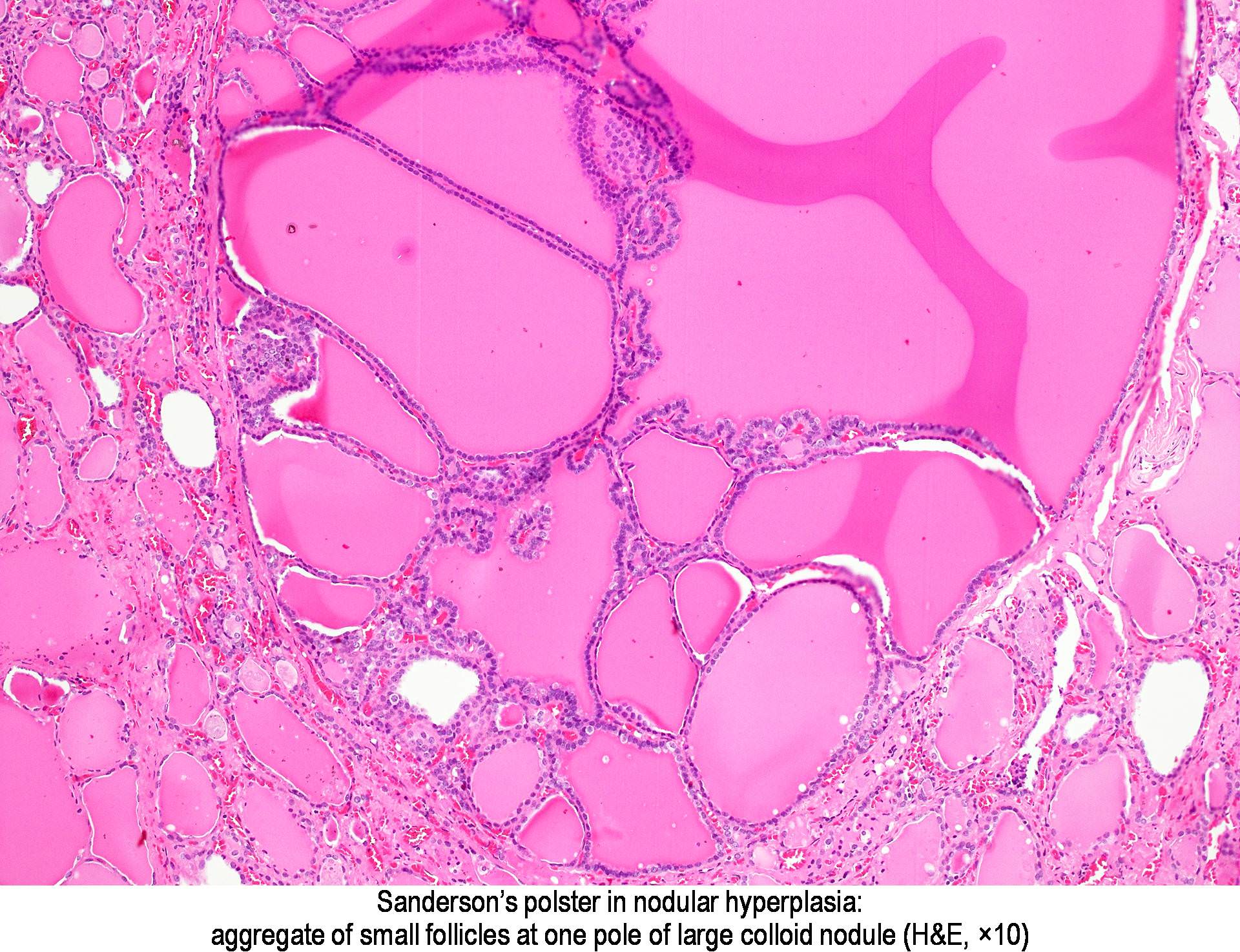
In shorts
- In normal thyroid gland or more commonly in hyperplastic conditions of thyroid gland such as nodular/multinodular goiter, adenomatoid goiter, or adenomatous hyperplasia, microscopically dilated thyroid follicles lined by cuboidal cells and filled with colloid may be seen.
- When colloid secretion increases in the small secondary follicles, their mass bulges into the follicular cavity and gives rise to a “Sanderson Polster” seen at one pole along the follicular wall of enlarged and dilated follicles.
- These dilated follicles are usually 2–3 times larger than the surrounding follicles
- Sanderson’s polster is composed mainly of small follicles lined by cuboidal cells
What is ‘Sanderson Polster’ ?
Sanderson polsters
When colloid secretion increases in the small secondary follicles, their mass bulges into the follicular cavity and gives rise to a
“Sanderson Polster” seen at one pole along the follicular wall of enlarged and dilated follicles
“Sanderson Polster” seen at one pole along the follicular wall of enlarged and dilated follicles
When colloid secretion increases in the small secondary follicles, their mass bulges into the follicular cavity and gives rise to a “Sanderson Polster” seen at one pole along the follicular wall of enlarged and dilated follicles.


