S1Q3T3 pattern MCQs
McGinn White sign
Key Points on S1Q3T3 Pattern (McGinn–White Sign)
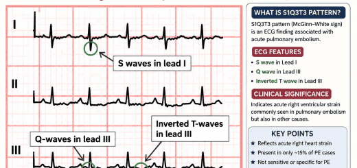
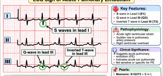
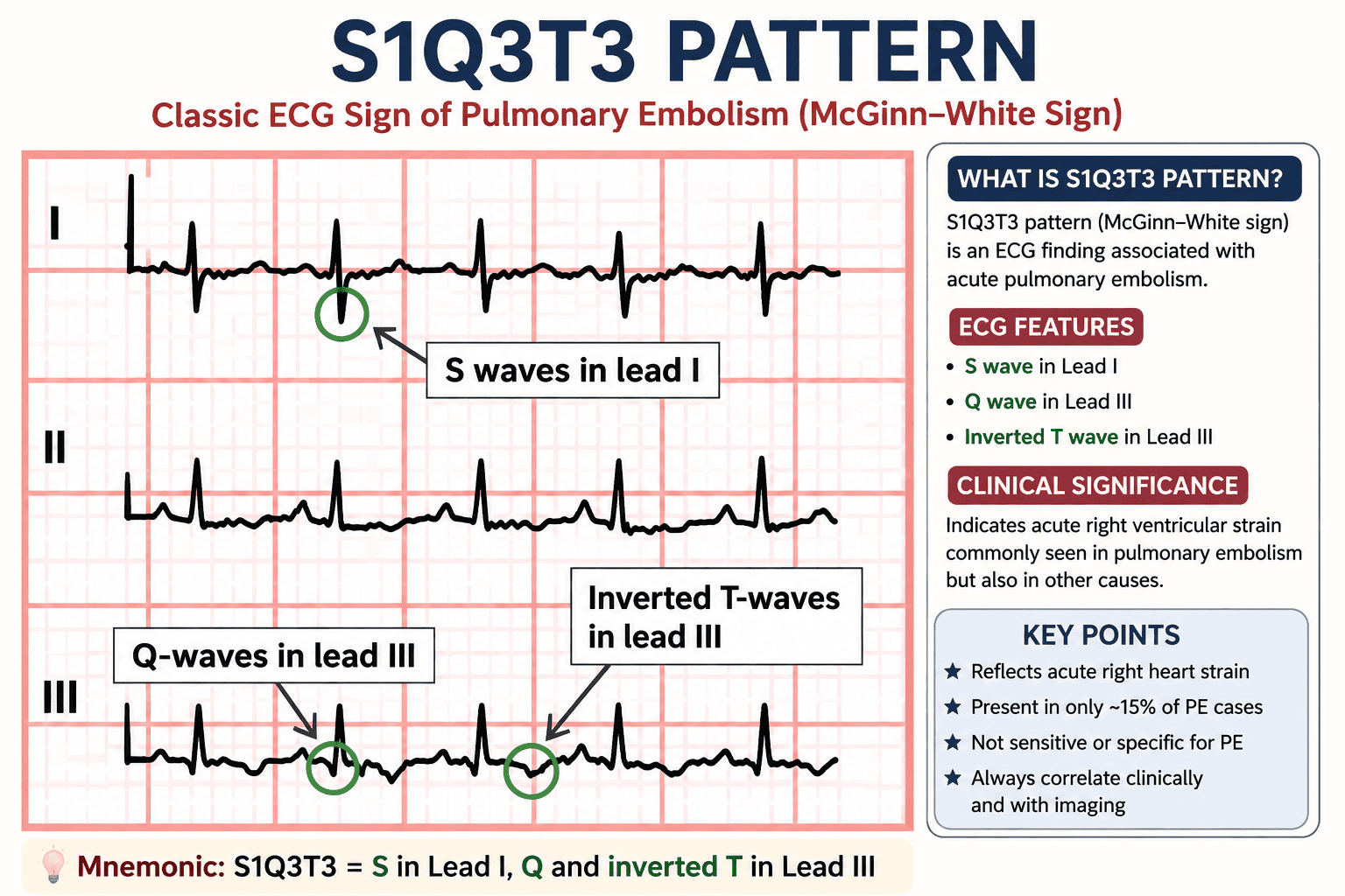
- S1Q3T3 pattern is a classic ECG sign of acute pulmonary embolism (PE), representing acute right ventricular (RV) strain rather than pulmonary embolism itself.
- S1Q3T3 stands for: S wave in Lead I, Q wave in Lead III, and T-wave inversion in Lead III.
- The S1Q3T3 pattern is also known as the McGinn–White Sign, first described by McGinn and White in 1935.
- Acute right ventricular pressure overload caused by pulmonary artery obstruction is the primary mechanism responsible for this ECG finding.
- The pattern occurs due to sudden elevation of pulmonary artery pressure, leading to right ventricular dilation and electrical axis deviation.
- Only about 15–20% of patients with acute pulmonary embolism demonstrate the classic S1Q3T3 pattern, making it a relatively uncommon finding.
- Sinus tachycardia is the most common ECG abnormality in pulmonary embolism, occurring more frequently than S1Q3T3.
- S1Q3T3 has low sensitivity and low specificity and therefore cannot confirm or exclude pulmonary embolism.
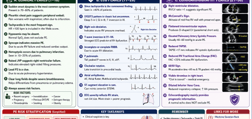
- Other ECG findings suggesting right ventricular strain include right axis deviation, incomplete or complete right bundle branch block (RBBB), and T-wave inversion in leads V1–V4.
- Anterior T-wave inversion (V1–V4) combined with S1Q3T3 may indicate a larger clot burden and more severe right ventricular dysfunction.
- S1Q3T3 is not exclusive to pulmonary embolism and may also occur in severe pulmonary hypertension, COPD exacerbation, acute asthma, ARDS, tension pneumothorax, and acute cor pulmonale.
- Patients with massive pulmonary embolism often present with hypotension, tachycardia, hypoxia, elevated jugular venous pressure, and evidence of RV strain on ECG.
- CT Pulmonary Angiography (CTPA) remains the gold-standard imaging test for diagnosing pulmonary embolism in hemodynamically stable patients.
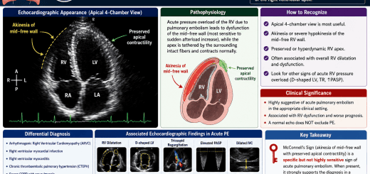
- Bedside echocardiography can rapidly identify right ventricular dilation, McConnell’s sign, and elevated pulmonary pressures in unstable patients.
- D-dimer testing is useful for excluding pulmonary embolism in low-risk patients but should always be interpreted alongside clinical probability scores.
- Clinical assessment using the Wells Score or Geneva Score helps determine the pre-test probability of pulmonary embolism before imaging.
- Treatment of pulmonary embolism includes anticoagulation, while systemic thrombolysis or catheter-directed therapy is reserved for high-risk or massive pulmonary embolism.
- Early recognition of the S1Q3T3 pattern may prompt timely evaluation for pulmonary embolism, reducing delays in diagnosis and improving patient outcomes.
- The absence of an S1Q3T3 pattern does not rule out pulmonary embolism, as many patients have a normal ECG or only nonspecific abnormalities.
- High-Yield Exam Pearl: S1Q3T3 = Acute Right Ventricular Strain Until Proven Otherwise. Always interpret the ECG together with the patient’s symptoms, risk factors, cardiac biomarkers, echocardiography, and CT pulmonary angiography.
1. S1Q3T3 pattern is classically associated with:
Answer: Pulmonary embolism
S1Q3T3 (McGinn–White sign) is a classic ECG sign of acute pulmonary embolism due to acute right ventricular strain. Although famous, it is neither sensitive nor specific.
S1Q3T3 (McGinn–White sign) is a classic ECG sign of acute pulmonary embolism due to acute right ventricular strain. Although famous, it is neither sensitive nor specific.
2. The “S1” in S1Q3T3 refers to:
Answer: Prominent S wave in Lead I
The S wave in Lead I results from rightward deviation of the QRS axis caused by acute right ventricular pressure overload.
The S wave in Lead I results from rightward deviation of the QRS axis caused by acute right ventricular pressure overload.
3. The “Q3” component indicates:
Answer: Q wave in Lead III
A small Q wave develops in Lead III due to altered ventricular depolarization from acute right ventricular strain.
A small Q wave develops in Lead III due to altered ventricular depolarization from acute right ventricular strain.
4. The “T3” component represents:
Answer: T-wave inversion in Lead III
T-wave inversion reflects acute right ventricular strain and ischemia secondary to pulmonary embolism.
T-wave inversion reflects acute right ventricular strain and ischemia secondary to pulmonary embolism.
5. Which ECG finding is MOST common in acute pulmonary embolism?
Answer: Sinus tachycardia
Sinus tachycardia is the most frequent ECG abnormality in pulmonary embolism. S1Q3T3 occurs in only a minority of patients.
Sinus tachycardia is the most frequent ECG abnormality in pulmonary embolism. S1Q3T3 occurs in only a minority of patients.
6. S1Q3T3 pattern primarily indicates:
Answer: Acute right ventricular strain
S1Q3T3 reflects acute right ventricular pressure overload. Although classically associated with pulmonary embolism, it simply indicates acute right heart strain.
S1Q3T3 reflects acute right ventricular pressure overload. Although classically associated with pulmonary embolism, it simply indicates acute right heart strain.
7. Approximately what percentage of patients with pulmonary embolism demonstrate the classic S1Q3T3 pattern?
Answer: 15–20%
Only a minority of pulmonary embolism patients show the classic S1Q3T3 pattern. Therefore, its absence does not exclude pulmonary embolism.
Only a minority of pulmonary embolism patients show the classic S1Q3T3 pattern. Therefore, its absence does not exclude pulmonary embolism.
8. The S1Q3T3 pattern was first described by:
Answer: McGinn and White
The ECG pattern was described in 1935 by Samuel McGinn and Paul Dudley White and is therefore known as the McGinn–White sign.
The ECG pattern was described in 1935 by Samuel McGinn and Paul Dudley White and is therefore known as the McGinn–White sign.
9. Which ECG finding frequently accompanies S1Q3T3 in acute pulmonary embolism?
Answer: Right bundle branch block
Right bundle branch block, right axis deviation, and anterior T-wave inversions commonly accompany acute right ventricular strain caused by pulmonary embolism.
Right bundle branch block, right axis deviation, and anterior T-wave inversions commonly accompany acute right ventricular strain caused by pulmonary embolism.
10. Which investigation is considered the imaging test of choice for confirming pulmonary embolism in a hemodynamically stable patient?
Answer: CT Pulmonary Angiography (CTPA)
CT pulmonary angiography is the gold-standard imaging test for diagnosing pulmonary embolism in most hemodynamically stable patients. ECG findings such as S1Q3T3 support the diagnosis but are never diagnostic by themselves.
CT pulmonary angiography is the gold-standard imaging test for diagnosing pulmonary embolism in most hemodynamically stable patients. ECG findings such as S1Q3T3 support the diagnosis but are never diagnostic by themselves.
11. Which of the following is NOT a component of the S1Q3T3 pattern?
Answer: ST elevation in Lead III
The classic S1Q3T3 pattern consists of a prominent S wave in Lead I, a Q wave in Lead III, and an inverted T wave in Lead III. ST-segment elevation is not part of this pattern.
The classic S1Q3T3 pattern consists of a prominent S wave in Lead I, a Q wave in Lead III, and an inverted T wave in Lead III. ST-segment elevation is not part of this pattern.
12. The primary mechanism responsible for the S1Q3T3 pattern is:
Answer: Acute increase in pulmonary vascular resistance causing RV strain
Pulmonary embolism suddenly elevates pulmonary artery pressure, producing acute right ventricular pressure overload and the characteristic ECG changes.
Pulmonary embolism suddenly elevates pulmonary artery pressure, producing acute right ventricular pressure overload and the characteristic ECG changes.
13. Which ECG finding is considered a marker of significant right ventricular strain in pulmonary embolism?
Answer: T-wave inversion in leads V1–V4
Anterior precordial T-wave inversion reflects right ventricular strain and is associated with larger pulmonary emboli and worse prognosis.
Anterior precordial T-wave inversion reflects right ventricular strain and is associated with larger pulmonary emboli and worse prognosis.
14. Which condition can also produce an S1Q3T3 pattern?
Answer: Tension pneumothorax
Any condition causing acute right ventricular pressure overload—including tension pneumothorax, severe asthma, COPD exacerbation, or pulmonary hypertension—may produce an S1Q3T3 pattern.
Any condition causing acute right ventricular pressure overload—including tension pneumothorax, severe asthma, COPD exacerbation, or pulmonary hypertension—may produce an S1Q3T3 pattern.
15. Which statement regarding the S1Q3T3 pattern is TRUE?
Answer: It is suggestive of acute right heart strain but is neither sensitive nor specific for pulmonary embolism.
The S1Q3T3 pattern is a classic but uncommon ECG sign. It should always be interpreted alongside the patient’s symptoms, clinical probability, biomarkers, and imaging studies such as CT pulmonary angiography.
The S1Q3T3 pattern is a classic but uncommon ECG sign. It should always be interpreted alongside the patient’s symptoms, clinical probability, biomarkers, and imaging studies such as CT pulmonary angiography.
16. The presence of an S1Q3T3 pattern should prompt evaluation for:
Answer: Pulmonary embolism with right ventricular strain
Although not diagnostic, S1Q3T3 should raise suspicion for pulmonary embolism, particularly in patients with acute dyspnea, pleuritic chest pain, tachycardia, hypoxia, or hemodynamic instability.
Although not diagnostic, S1Q3T3 should raise suspicion for pulmonary embolism, particularly in patients with acute dyspnea, pleuritic chest pain, tachycardia, hypoxia, or hemodynamic instability.
17. Which of the following ECG findings is commonly associated with a large pulmonary embolism?
Answer: Right axis deviation
Acute right ventricular pressure overload frequently produces right axis deviation. Other associated findings include RBBB, anterior T-wave inversion, and the S1Q3T3 pattern.
Acute right ventricular pressure overload frequently produces right axis deviation. Other associated findings include RBBB, anterior T-wave inversion, and the S1Q3T3 pattern.
18. In a patient with suspected pulmonary embolism, S1Q3T3 should be interpreted as:
Answer: A supportive ECG finding that requires clinical correlation and imaging
The diagnosis of pulmonary embolism is based on clinical probability combined with investigations such as D-dimer, CT pulmonary angiography, echocardiography, or ventilation–perfusion scanning.
The diagnosis of pulmonary embolism is based on clinical probability combined with investigations such as D-dimer, CT pulmonary angiography, echocardiography, or ventilation–perfusion scanning.
19. Acute pulmonary embolism causes S1Q3T3 primarily because of:
Answer: Sudden pressure overload of the right ventricle
Pulmonary artery obstruction abruptly increases pulmonary vascular resistance, leading to acute right ventricular dilation, pressure overload, and characteristic ECG manifestations.
Pulmonary artery obstruction abruptly increases pulmonary vascular resistance, leading to acute right ventricular dilation, pressure overload, and characteristic ECG manifestations.
20. Which of the following statements BEST summarizes the clinical value of the S1Q3T3 pattern?
Answer: It is a classic but uncommon sign of acute right ventricular strain and should always be interpreted with the clinical picture.
The S1Q3T3 (McGinn–White) pattern is a valuable examination and teaching point but has low sensitivity and specificity for pulmonary embolism. It supports the diagnosis when combined with clinical findings, biomarkers, echocardiography, and CT pulmonary angiography.
The S1Q3T3 (McGinn–White) pattern is a valuable examination and teaching point but has low sensitivity and specificity for pulmonary embolism. It supports the diagnosis when combined with clinical findings, biomarkers, echocardiography, and CT pulmonary angiography.