Image Question-47
What is the diagnosis of IMAGE?
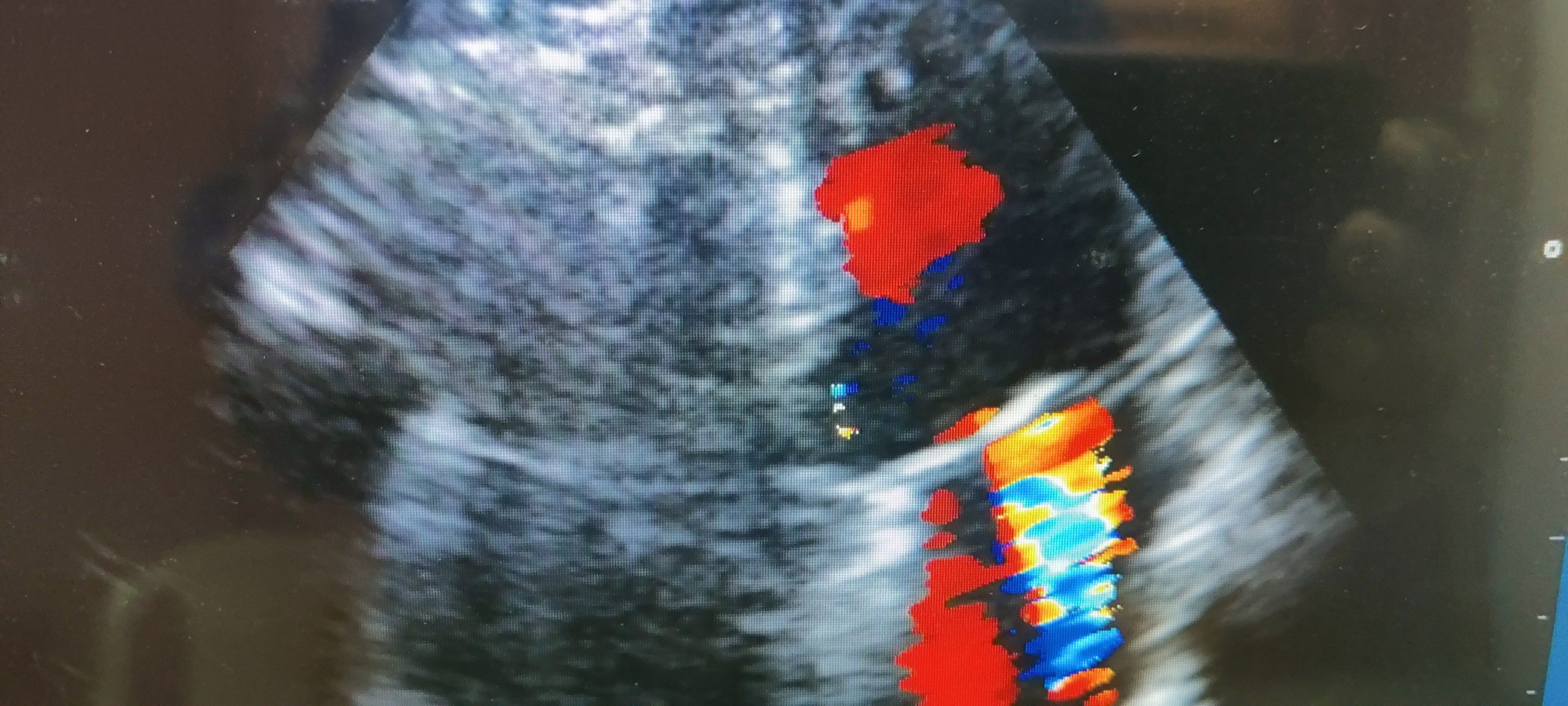
A. Aortic Regurgitation
B. Mitral Regurgitation
C. Mitral Stenosis
D. Aortic Stenosis
Mitral Regurgitation – Indicators of Severity
• Mitral valve pathology
• LV/ LA size
• Color Doppler: – Vena contracta, Jet Area, Flow convergence
• Mitral E; Pulmonary vein pattern
• Regurgitant flow/fraction
• CW – density and contour
Etiology and mechanism of mitral regurgitation
| Etiology of mitral regurgitation | Mechanism of mitral regurgitation |
|---|---|
| Atrial fibrillation | Annular dilation, leaflet mal-coaptation |
| Acute ischemia | Papillary muscle dysfunction or rupture |
| Congenital or genetic disorders; Marfan syndrome, Ehlers-Danlos syndrome, Down syndrome | Leaflet prolapse, cleft or rudimentary leaflets |
| Endocarditis; infective and marantic | Leaflet perforation, mal-coaptation, chordal rupture |
| Drugs; fenfluramine and dexfenfluramine | Leaflets, chordae |
| Functional/secondary; dilated cardiomyopathy | Left ventricular remolding, papillary muscle displacement leading to leaflet tethering and annulus dilation |
| Hypertrophic obstructive cardiomyopathy | Systolic anterior motion of anterior mitral valve leaflet |
| Myxomatous degeneration (primary) (1) Barlow’s disease (2) Fibroelastic deficiency | Leaflets prolapse Rupture chordae |
| Mitral annular calcifications | Annulus, leaflets |
| Rheumatic heart disease | Leaflets, chordae |
| Radiation | Leaflets, chordae |
Grading the severity of mitral regurgitation
| Mild | Moderate | Severe | |
|---|---|---|---|
| Qualitative parameters | |||
| MV morphology | Normal/abnormal | Normal/abnormal | Flail leaflet/chordal rupture |
| Color flow Doppler of MR jet* | < 20% of LA size | 20%-40% of LA size | > 40% of LA size |
| Continuous wave Doppler MR jet density MR jet contour | Faint Parabolic | Dense Parabolic | Dense Early peaking-triangular |
| Flow convergence zone* | No or small | Intermediate | Large |
| Semi-quantitative parameters | |||
| Vena contracta | < 0.3 cm | 0.3-0.69 cm | ≥ 0.7 cm |
| Mitral valve inflow | A-wave dominant | E-wave dominant, > 1.2 m/s | |
| Mitral to aortic TVI ratio < 1 m/s | Mitral to aortic TVI ratio 1 to 1.4 m/s | Mitral to aortic TVI > 1.4 m/s | |
| Pulmonary veins flow | Systolic dominance | Normal or systolic blunting | Systolic flow reversal in > 1 vein |
| LA/LV size | Normal | Intermediate | Enlarged, particularly with normal LV function |
| Quantitative parameters | |||
| Effective regurgitant orifice area by PISA or 3D color Doppler echo | < 0.2 cm2 | 0.2-0.29 cm2; Mild to moderate 0.3-0.39 cm2; Moderate to severe | ≥ 0.4 cm2 |
| Regurgitant volume | < 30 mL/beat | 30-44 mL/beat; Mild to moderate 45-59 mL/beat; Moderate to severe | ≥ 60 mL/beat |
| Regurgitant fraction | < 30% | 30%-39%; Mild to moderate 40%-49%; Moderate to severe | ≥ 50% |
MR: mitral regurgitation; MV: mitral valve; LA: left atrium; LV: left ventricle; TVI: time velocity integral. *At Nyquist limit between 50-70 cm/s. Color Doppler gain needs to be optimized


