De Winter Sign (a STEMI-equivalent ECG pattern)
De Winter Sign (a STEMI-equivalent ECG pattern)
| No. | De Winter sign – Advanced Clinical Point |
|---|---|
| 1 | De Winter sign is a STEMI equivalent pattern seen on ECG. |
| 2 | It reflects acute occlusion of the proximal left anterior descending (LAD) coronary artery. |
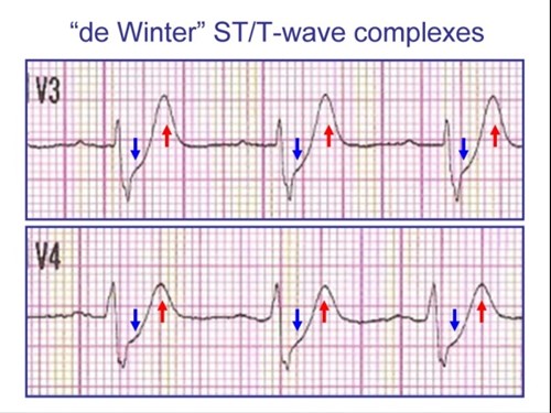
| 3 | Characterized by upsloping ST depression at the J-point in precordial leads (V1–V4). |
| 4 | Accompanied by tall, symmetrical, and peaked T waves in the same leads. |
| 5 | ST elevation is notably absent in anterior leads, making it easy to miss. |
| 6 | May show mild ST elevation (0.5–1 mm) in lead aVR. |
| 7 | Represents ongoing transmural ischemia, similar in severity to classic STEMI. |
| 8 | Accounts for approximately 2–3% of anterior myocardial infarctions. |
| 9 | Often misinterpreted due to absence of ST elevation and resemblance to non-specific ST/T changes. |
| 10 | Unlike hyperkalemia, the tall T waves are broad-based and symmetric. |
| 11 | It is a static ECG pattern—does not typically evolve like classic STEMI unless reperfusion occurs. |
| 12 | QRS duration may be normal or slightly widened, depending on the extent of ischemia. |
| 13 | May be associated with reciprocal ST changes in inferior leads (e.g., II, III, aVF). |
| 14 | Patients often present with acute chest pain, similar to STEMI presentations. |
| 15 | Recognition mandates immediate reperfusion therapy—usually urgent PCI. |
| 16 | Not associated with ST elevation in V1–V4, making it different from traditional anterior STEMI. |
| 17 | Requires differentiation from Wellens syndrome, which has different T-wave evolution and occurs post-ischemia. |
| 18 | Failure to identify De Winter sign may delay lifesaving treatment. |
| 19 | De Winter sign patients have similar mortality and morbidity outcomes as STEMI if not promptly treated. |
| 20 | De Winter pattern is now officially recognized as a STEMI equivalent in international guidelines (ACC/AHA/ESC). |
ECG Diagnostic Criteria – De Winter Sign
| Criteria | Description |
|---|---|
| T Waves | Tall, prominent, symmetrical T waves in the precordial leads (V1–V4) |
| ST Depression | Upsloping ST segment depression > 1 mm at the J point in the precordial leads |
| ST Elevation in Precordial Leads | Not present (absence of typical ST elevation in V1–V4) |
| aVR Changes | Mild reciprocal ST elevation (0.5 mm – 1 mm) may be seen in lead aVR |
| STEMI Evolution | Typical STEMI morphology may precede or follow the De Winter pattern |
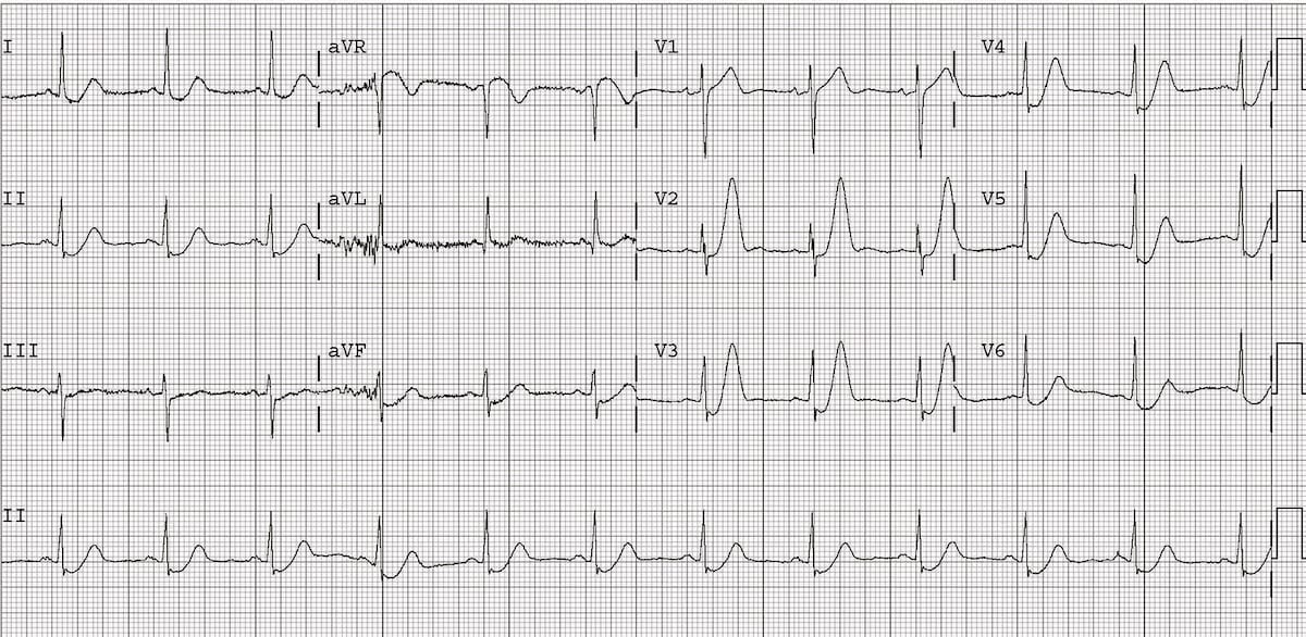
De Winter T waves
Upsloping ST depression (> 1mm at J point) in the precordial leads V2-6, plus leads I and II
Peaked anterior T waves, with the ascending limb of the T wave commencing below the isoelectric baseline
ST elevation in aVR > 0.5mm


