Circle of Willis
🧠 Circle of Willis – MCQs
🧠 Circle of Willis – Key Summary
| Artery | Origin | Connections | Clinical Notes |
|---|---|---|---|
| Anterior cerebral artery (ACA) | Internal carotid artery | Connected to opposite ACA via Anterior Communicating Artery | Supplies medial frontal & parietal lobes |
| Anterior communicating artery (ACoA) | Single vessel | Joins left and right ACA | Most common site of berry aneurysm |
| Middle cerebral artery (MCA) | Internal carotid artery | Not part of the circle; runs laterally | Main supplier of lateral cerebral hemispheres |
| Internal carotid artery (ICA) | Common carotid artery | Gives rise to ACA, MCA, and PCoA | Traverses cavernous sinus before Circle |
| Posterior communicating artery (PCoA) | Internal carotid artery | Connects ICA to Posterior Cerebral Artery | Frequently hypoplastic or absent |
| Posterior cerebral artery (PCA) | Basilar artery | Connected to ICA via PCoA | Supplies occipital lobe (visual cortex) |
| Basilar artery | Union of vertebral arteries | Terminates as PCAs | Runs along clivus; can be compressed by tumors |
| Vertebral arteries | Subclavian arteries | Join to form basilar artery | Enter skull via foramen magnum |
| Component | Source Vessel | Connection/Branching | Clinical Relevance |
|---|---|---|---|
| Anterior cerebral artery (ACA) | Internal carotid artery (ICA) | Connected to contralateral ACA via anterior communicating artery (ACoA) | Occlusion → contralateral leg weakness, sensory loss |
| Anterior communicating artery (ACoA) | Connects left & right ACA | Completes anterior part of circle | Common site of berry aneurysm |
| Internal carotid artery (ICA) | Common carotid artery | Gives rise to ACA, MCA, and PCoA | ICA stenosis → TIA/stroke; site for carotid endarterectomy |
| Middle cerebral artery (MCA) | ICA (not part of circle) | Lateral cerebral hemisphere | Most common artery involved in ischemic stroke |
| Posterior communicating artery (PCoA) | ICA | Connects ICA with PCA | Frequent aneurysm site (causing CN III palsy) |
| Posterior cerebral artery (PCA) | Basilar artery (terminal branch) | Supplies occipital lobe, thalamus, midbrain | Occlusion → contralateral homonymous hemianopia |
| Basilar artery | Union of vertebral arteries | Terminates as left & right PCA | Basilar thrombosis → devastating brainstem stroke |
| Vertebral arteries | Subclavian arteries | Join to form basilar artery | Vertebral dissection → posterior circulation stroke |
The circle of Willis is a ring of interconnected arteries located at the base of the brain. It connects the brain’s major arterial systems—the internal carotid arteries and the vertebrobasilar system—providing a critical backup pathway for blood flow. If a vessel becomes blocked or narrowed, the circle can reroute blood, which may prevent an ischemic stroke or reduce its severity.
Arteries that form the circle of Willis
The circle is a polygon-shaped structure created by the following arteries:
- Anterior cerebral arteries (left and right): Branches of the internal carotid arteries that supply the majority of the frontal and superior parietal lobes. They form the front of the circle.
- Anterior communicating artery: A short, midline vessel that connects the two anterior cerebral arteries.
- Internal carotid arteries (left and right): Though they do not technically form the circle, they feed into it by branching into the anterior and middle cerebral arteries.
- Posterior cerebral arteries (left and right): Terminal branches of the basilar artery that supply the occipital lobe and parts of the temporal lobe. They form the back of the circle.
- Posterior communicating arteries (left and right): Connect the internal carotid arteries and the posterior cerebral arteries, completing the ring.
Function and variations
Collateral circulation
The primary purpose of the circle of Willis is to provide collateral circulation, or a backup blood supply, to the brain.
- If a major artery leading to the brain is compromised, blood can be rerouted from the other vessels in the circle to ensure the brain continues to receive oxygen.
- However, this compensatory effect is not guaranteed. Many people have anatomical variations where certain vessels are absent, duplicated, or smaller in caliber (hypoplastic).
- Studies suggest that a complete, or “classic,” circle of Willis is found in less than half of the population.
Common variants
Variations are common and do not necessarily cause problems, but they can affect how the brain compensates during a vascular event. Common variations include:
- Fenestration: A single vessel divides into two separate channels before joining back together.
- Hypoplastic vessels: One of the connecting arteries is significantly narrower than normal.
- Incomplete posterior communicating arteries: The most common variation, where one or both posterior communicating arteries are underdeveloped or missing.
Clinical significance
Anomalies of the circle of Willis are clinically important, as they are associated with several cerebrovascular conditions.
- Aneurysms: The junctions within the circle of Willis are common sites for cerebral aneurysms, which are weakened, bulging areas in an artery wall. Ruptured aneurysms can cause a life-threatening brain hemorrhage.
- Stroke risk: While a complete circle provides some protection against stroke, variations like hypoplastic vessels can increase the risk of a more severe ischemic stroke.
- Moyamoya disease: A rare chronic condition involving progressive narrowing of the arteries within and around the circle of Willis.
- Surgery: The circle of Willis and surrounding structures are at risk of damage during surgery at the base of the brain, particularly procedures involving aneurysms or the pituitary gland.
Which of the following is not a part of Circle of Willis
[A] Basilar artery
[B] Vertebral artery
[C] Anterior communicating artery
[D] Anterior cerebral arteries
Most commonly involved artery in fenestrations and duplications as physiologic variant in circle of willis is
[A] Basilar artery
[B] Middle cerebral artery
[C] Anterior communicating artery
[D] Anterior cerebral arteries
Subclavian steal syndrome cause
[A] Reduced perfusion to Brain
[B] Increased perfusion to Brain
[C] Reduced perfusion to ipsilateral upper limb
[D] Reduced perfusion to contralateral upper limb
Subclavian steal syndrome usually block seen in
[A] proximal stenosis of subclavian artery
[B] distal stenosis of subclavian artery
[C] proximal stenosis of vertebral artery
[D] distal stenosis of vertebral artery
Most common locations for intracranial aneurysms
[A] Basilar artery
[B] Putamen
[C] Anterior communicating artery
[D] Midbrain
Circle of Willis – composed of the following arteries:
- Anterior cerebral artery (left and right) at their A1 segments
- Anterior communicating artery
- Internal carotid artery (left and right) at its distal tip (carotid terminus)
- Posterior cerebral artery (left and right) at their P1 segments
- Posterior communicating artery (left and right)

Subclavian steal syndrome
- In subclavian steal syndrome – blood is “stolen” from the vertebral artery on the affected side to preserve blood flow to the upper limb.
- Subclavian steal syndrome results from a proximal stenosis of the subclavian artery
The Circle of Willis is a ring-shaped arterial structure at the base of the brain that provides collateral blood flow between the anterior and posterior cerebral circulations. It is an important safety mechanism that helps maintain cerebral perfusion if one major vessel is blocked or narrowed.
🔹 Anatomy of the Circle of Willis
It forms a polygonal anastomotic system and usually includes:
Anterior Circulation (from Internal Carotid Arteries):
- Anterior cerebral arteries (ACA) – paired, connected by the anterior communicating artery.
- Anterior communicating artery (ACoA) – single vessel connecting the two ACAs.
- Internal carotid arteries (ICA) – contribute to the middle cerebral arteries (not part of the circle but arise here) and posterior communicating arteries.
Posterior Circulation (from Vertebrobasilar System):
- Posterior cerebral arteries (PCA) – paired, terminal branches of the basilar artery.
- Posterior communicating arteries (PCoA) – paired, connecting ICA to PCA.
- Basilar artery – formed by union of the two vertebral arteries.
🔹 Configuration
- Complete Circle: Found in ~34–50% of people.
- Variations: Hypoplasia, absence, or asymmetry of one or more communicating arteries are very common.
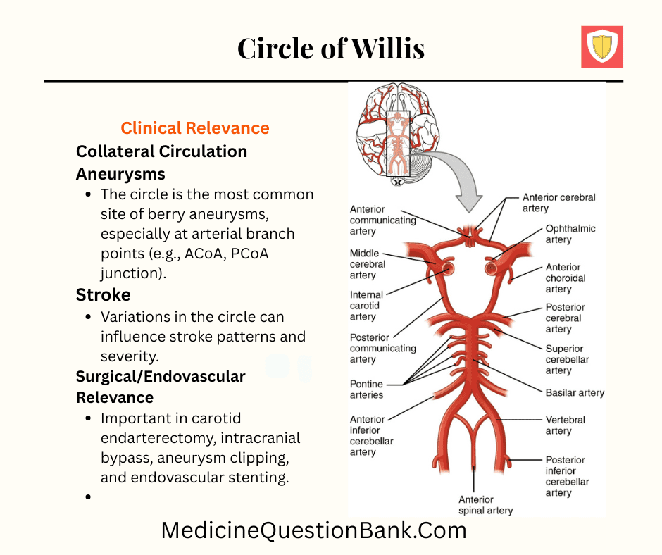
🔹 Clinical Importance
- Collateral Circulation
- If one artery is blocked (e.g., ICA stenosis), the circle can maintain cerebral perfusion through alternative pathways.
- Aneurysms
- The circle is the most common site of berry aneurysms, especially at arterial branch points (e.g., ACoA, PCoA junction).
- Stroke
- Variations in the circle can influence stroke patterns and severity.
- Surgical/Endovascular Relevance
- Important in carotid endarterectomy, intracranial bypass, aneurysm clipping, and endovascular stenting.


