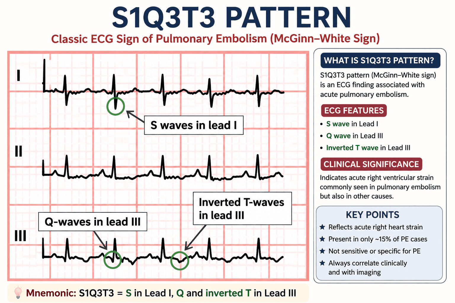
S1Q3T3 pattern
S1Q3T3 Pattern (McGinn–White Sign): A Comprehensive Guide
S1Q3T3 Pattern (McGinn–White Sign): Summary Table
| Feature | Description |
|---|---|
| Definition | Classic ECG pattern associated with acute pulmonary embolism (PE) due to acute right ventricular (RV) strain. |
| Also Known As | McGinn–White Sign |
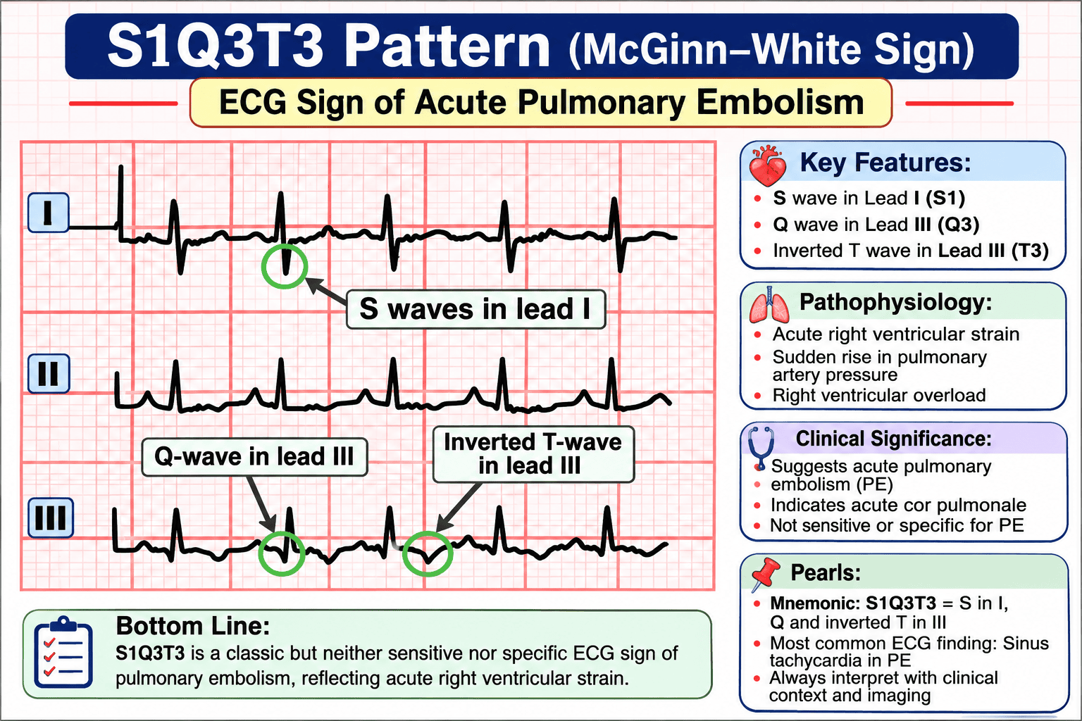
| Mnemonic | S1Q3T3 = S wave in Lead I + Q wave in Lead III + T-wave inversion in Lead III |
| S1 | Prominent S wave in Lead I indicating rightward shift of the QRS axis. |
| Q3 | Small Q wave in Lead III caused by altered ventricular depolarization. |
| T3 | Inverted T wave in Lead III reflecting right ventricular strain. |
| Underlying Mechanism | Acute obstruction of pulmonary arteries → Increased pulmonary vascular resistance → RV pressure overload → Right ventricular dilation and strain. |
| Most Common Cause | Acute pulmonary embolism (PE). |
| Other Causes | Severe pulmonary hypertension, acute cor pulmonale, COPD exacerbation, severe asthma, ARDS, tension pneumothorax, extensive pneumonia. |
| Sensitivity for PE | Approximately 15–20% (low sensitivity). |
| Specificity | Low; not specific for pulmonary embolism. |
| Most Common ECG Finding in PE | Sinus tachycardia. |
| Associated ECG Findings | Right axis deviation, right bundle branch block (RBBB), P pulmonale, T-wave inversion in V1–V4, clockwise rotation. |
| Clinical Significance | Indicates acute right ventricular strain rather than pulmonary embolism itself. |
| Prognostic Value | May indicate large clot burden and severe RV dysfunction when accompanied by hypotension, RBBB, and anterior T-wave inversions. |
| Diagnosis of PE | Requires clinical assessment, Wells score, D-dimer (when appropriate), CT pulmonary angiography (CTPA), echocardiography in unstable patients, or V/Q scan. |
| Treatment | Depends on PE severity: anticoagulation, thrombolysis, catheter-directed therapy, or surgical embolectomy. |
| Exam Pearl | S1Q3T3 is a classic but uncommon ECG sign of PE and should never be used alone to diagnose pulmonary embolism. |
| High-Yield Take-Home Message | S1Q3T3 = Acute Right Heart Strain Until Proven Otherwise. Always correlate with clinical findings and imaging. |

What is the S1Q3T3 Pattern?
The S1Q3T3 pattern, also known as the McGinn–White sign, is a classic electrocardiographic (ECG) finding associated with acute pulmonary embolism (PE). It reflects acute right ventricular (RV) strain due to a sudden increase in pulmonary vascular resistance.
The pattern consists of:
- S1 – Prominent S wave in Lead I
- Q3 – Q wave in Lead III
- T3 – Inverted T wave in Lead III
It was first described by Samuel McGinn and Paul Dudley White in 1935. (PubMed Central (PMC))
ECG Features
S wave in Lead I
- Indicates right axis deviation
- Caused by acute RV overload
Q wave in Lead III
- Small but distinct Q wave
- Represents altered ventricular depolarization
T-wave inversion in Lead III
- Reflects RV ischemia/strain
- Often accompanies pulmonary hypertension
Why Does It Occur?
Pulmonary embolism causes:
- Sudden obstruction of pulmonary arteries
- Acute rise in pulmonary artery pressure
- Right ventricular dilation
- Right ventricular strain
- Rightward shift of the electrical axis
These changes produce the characteristic ECG pattern.
Clinical Significance
S1Q3T3 suggests:
- Acute pulmonary embolism
- Acute cor pulmonale
- Significant right ventricular strain
However:
It is neither sensitive nor specific for pulmonary embolism.
Most patients with PE do not show S1Q3T3, and the pattern can occur in several other causes of acute RV strain. (PubMed Central (PMC))
Sensitivity
Recent systematic reviews estimate the S1Q3T3 pattern is present in about 15% of confirmed acute pulmonary embolism cases. (PubMed Central (PMC))
More Common ECG Findings in Pulmonary Embolism
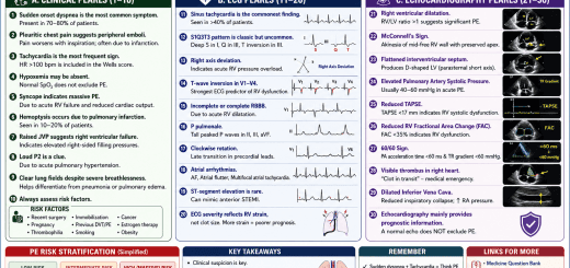
- Sinus tachycardia (most common)
- Right axis deviation
- Right bundle branch block
- T-wave inversion in V1–V4
- P pulmonale
- Clockwise rotation
- Atrial arrhythmias
- S1Q3T3 pattern
Differential Diagnosis
S1Q3T3 may also occur in:
- Severe pulmonary hypertension
- Acute severe asthma
- COPD exacerbation
- Tension pneumothorax
- Massive pneumonia
- Acute respiratory distress syndrome (ARDS)
- Acute cor pulmonale
Prognostic Importance
When accompanied by:
- RV dysfunction
- Right precordial T-wave inversion
- RBBB
- Hypotension
the ECG suggests a larger clot burden and higher-risk pulmonary embolism. (PubMed Central (PMC))
Diagnosis of Pulmonary Embolism
S1Q3T3 alone does not diagnose PE.
Diagnosis should integrate:
- Clinical probability (e.g., Wells score)
- D-dimer (when appropriate)
- CT pulmonary angiography (CTPA)
- Echocardiography in unstable patients
- Ventilation–perfusion (V/Q) scan when indicated
High-Yield NEET-SS / DM Cardiology Pearls
- Mnemonic: S1Q3T3 = S in Lead I, Q and inverted T in Lead III
- Represents acute right heart strain, not PE itself.
- Sinus tachycardia is the most common ECG abnormality in PE.
- S1Q3T3 is classically associated with PE but has low sensitivity and specificity.
- Right precordial T-wave inversions (V1–V4) and RBBB often indicate more severe RV strain.
- ECG findings should always be interpreted in the context of the patient’s symptoms and imaging results. (PubMed Central (PMC))
SEO Meta Title
S1Q3T3 Pattern: ECG Sign of Pulmonary Embolism | Causes, Diagnosis & Clinical Significance
Meta Description
Learn about the S1Q3T3 (McGinn–White) ECG pattern, its association with pulmonary embolism, causes, diagnostic value, sensitivity, prognosis, and key exam pearls for medical students and cardiologists.
Focus Keywords
- S1Q3T3 pattern
- McGinn White sign
- S1Q3T3 ECG
- Pulmonary embolism ECG
- Right ventricular strain ECG
- ECG signs of pulmonary embolism
- Acute pulmonary embolism ECG
- S1Q3T3 meaning
- PE ECG findings
- Cardiology ECG patterns