Cardiology MCQs-7
Normal QRS axis is between
A. -30° and +60°
B. -60° and +90°
C. -30° and +120°
D. -30° and +90°
Most common cause of marked left axis deviation
A. LAFB
B. Endocardial cushion defects
C.Left ventricular hypertrophy
D. Inferior wall myocardial infarction
Basic requirements for diagnosis of LBBB include QRS duration of
A. 90 milliseconds or more
B. 120 milliseconds or more
C. 160 milliseconds or more
D. 180 milliseconds or more
In Left Bundle Branch Block Prolonged time to peak R wave in V5 and V6 is usually
A. >30 msec
B. >60 msec
C. >90 msec
D. >120 msec
In RBBB ‘S’ waves in leads I and V6 is usually
A. ≥ 20 msec wide
B. ≥ 40 msec wide
C. ≥ 60 msec wide
D. ≥ 75 msec wide
In RBBB time to peak R wave in leads V1
A. > 30 msec
B. > 50 msec
C. > 90 msec
D. Normal duration
Which criteria is used in the diagnosis of an acute myocardial infarction when a left bundle branch block is present?
A. Feighner Criteria
B. Amsterdam criteria
C. Sgarbossa criteria
D. McDonald criteria
Common Diagnostic Criteria for Bundle Branch Blocks
Complete Left Bundle Branch Block
- QRS duration ≥ 120 msec
- Broad, notched, or slurred R waves in leads I, aVL, V5, and V6
- Small or absent initial r waves in leads V1 and V2 followed by deep S waves
- Absent septal q waves in leads I, V5 , and V6
- Prolonged time to peak R wave (>60 msec) in V5 and V6
Complete Right Bundle Branch Block
- QRS duration ≥ 120 msec
- rsr′, rsR′, or rSR′, patterns in leads V1 and V2
- S waves in leads I and V6 ≥ 40 msec wide
- Normal time to peak R wave in leads V5 and V6 but > 50 msec in V1
LBBB on ECG – Diagnostic criteria are defined by the American College of Cardiology (ACC) and American Heart Association (AHA) as follows:
- Rhythm must be of super-ventricular origin (EG: ventricular activation coming from atrial or AV nodal activation)
- QRS Duration greater than 120 ms
- Lead V1 should have either a QS or a small r wave with large S wave
- Lead V6 should have a notched R wave and no Q wave
Incomplete LBBB
Incomplete LBBB may result from lesser degrees of conduction delay in the left bundle branch system.
Features include modest prolongation of the QRS complex (100 to 119 msec)
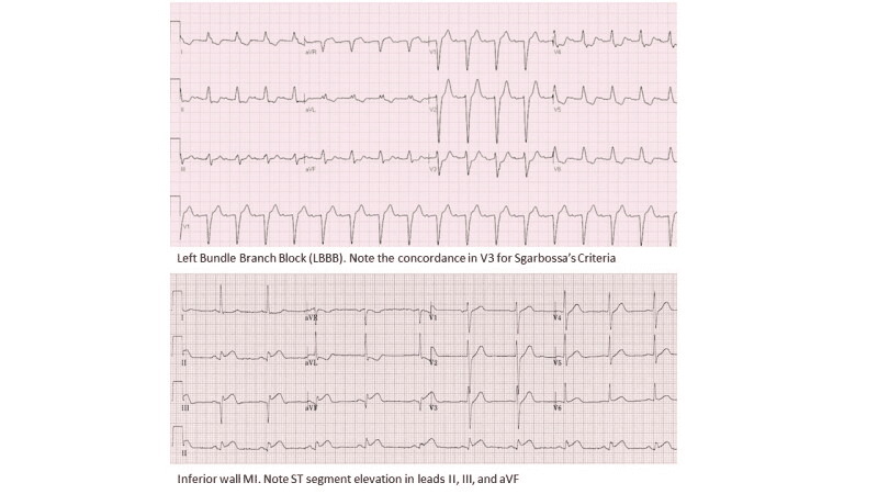
ECG- Left Bundle Branch Block and inferior wall MI.
Contributed by Steven Mountfort. Creative Commons Attribution-NonCommercial-NoDerivatives 4.0 International (CC BY-NC-ND 4.0) ( http://creativecommons.org/licenses/by-nc-nd/4.0/ )


