Emphysematous Cholecystitis
Emphysematous cholecystitis-
Begin with – acute cholecystitis (calculous or acalculous)
Followed by
- Ischemia or gangrene of the gallbladder wall
- Infection by gas-producing organisms.
Bacteria most frequently cultured
Most frequently cultured Bacteria –
Anaerobes- Clostridium welchii or C. perfringens
Aerobes – E. coli.
Risk Factors
Most frequently in elderly men and in patients with diabetes mellitus.
Diagnosis
During an ultrasonogram- air in the wall or lumen of the gallbladder can interfere with the clear visualization of the gallbladder
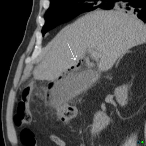
Best imaging modality to confirm the presence of emphysematous cholecystitis
The best imaging modality to confirm the presence of emphysematous cholecystitis is a contrast-enhanced abdominal CT scan.
The plain x-ray may reveal air and/or air-fluid levels in the gallbladder.
Plain abdominal film by finding gas within the gallbladder lumen, dissecting within the gallbladder wall to form a gaseous ring, or in the pericholecystic tissues.
Morbidity and Mortality rates
Pathology
Emphysematous cholecystitis shows a higher degree of endarteritis obliterans when compared to acute cholecystitis.
The causative organisms are E coli, Aerobactor aerogens, Klebsiella spp, and Salmonella spp.
Gangrene and perforation and pericholecystic abscess may ensue
The morbidity and mortality rates with emphysematous cholecystitis are High
Emphysematous cholecystitis occurs in about 1% of all cases of acute cholecystitis.
Characteristic feature
Characteristic feature of this sinister variant of cholecystitis is the presence of gas in the lumen and wall of the gallbladder
Classification according to radiological findings as progressive stages
Early stage – Air limited to the gallbladder lumen
Most advanced stage – Air in the gallbladder and the pericholecystic tissue
Management
Prompt surgical intervention and appropriate antibiotics
Micro-organisms that have been isolated in patients with emphysematous cholecystitis include the following:
- Clostridia
- Klebsiella
- Escherichia coli
- Enterococci
- Anaerobic streptococci
Emphysematous cholecystitis with local perforation