Image Question-56
What is the name of the wave marked by arrow?
[A] L wave
[B] M wave
[C] R wave
[D] Y wave
L-wave in Transmitral flow Doppler waves is a marker of
[A] Advanced diastolic dysfunction
[B] Advanced systolic dysfunction
[C] Improved diastolic dysfunction
[D] Improved systolic dysfunction
A patient 59 years male admitted to ICU with complains of breathlessness. Echocardiogram shows L-wave in Transmitral flow Doppler. Which of the following condition L-wave in Transmitral flow Doppler is not likely to disappear?
[A] Improved fluid management
[B] Improved heart function
[C] Decreased heart rate
[D] Revert to sinus rhythm
Transmitral flow Doppler waves
Transmitral flow Doppler waves, specifically the E and A waves, are used to assess left ventricular diastolic function by measuring the velocity of blood flow across the mitral valve during early and late diastole, respectively.
What are transmitral flow Doppler waves?
Doppler echocardiography is a non-invasive ultrasound technique that measures blood flow velocities.
Transmitral flow refers to the blood flow across the mitral valve, which separates the left atrium and left ventricle.
Doppler waves, specifically the E and A waves, are patterns of blood flow velocity during early and late diastole (the period between heartbeats when the ventricles are filling with blood).
What are the E and A waves?
E wave : Represents the rapid early filling of the left ventricle during diastole.
A wave : Represents the filling of the left ventricle during atrial contraction (atrial kick).
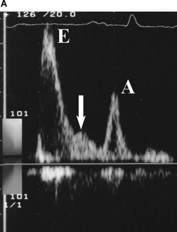
‘L’ wave in echocardiography
L wave in echocardiography refers to a mid-diastolic flow seen in pulse wave Doppler and M-mode echocardiography. It indicates continued pulmonary vein flow into the left ventricle across the left atrium
This phenomenon is significant in clinical settings as it can suggest advanced diastolic dysfunction and elevated left ventricular filling pressures, particularly in patients with conditions like left ventricular hypertrophy (LVH)
The presence of an L wave is associated with poorer prognosis and is used to predict future heart failure events
What is an ‘L’ wave?
An L wave is a mid-diastolic transmitral flow velocity that appears on echocardiography during the quiescent period of diastole, between early and late filling.
Why does ‘L’ wave appear?
The L wave is often associated with elevated left ventricular filling pressures and advanced diastolic dysfunction.
Why does ‘L’ wave disappear?
- Improved fluid management: Diuretics and other treatments aimed at reducing fluid overload can lead to a decrease in left ventricular filling pressures, which can cause the L wave to disappear.
- Improved heart function: As the heart’s ability to relax and fill properly improves, the L wave may also disappear.
- Increased heart rate: As heart rate increases, the diastolic filling phase is shortened, and the L wave may become absorbed into the E and A waves.
- Presence of A wave: In the condition with the presence of A wave, A wave occupies late diastolic phase and shortens the time for L wave existence.
- Clinical Significance:The presence of an L wave can be an indicator of diastolic dysfunction and elevated left ventricular filling pressures, while its disappearance can suggest successful treatment and improved heart function.
- Heart rhythm: The L wave can also be influenced by factors like heart rhythm (e.g., atrial fibrillation).
Types of L-waves.
Two types of L-waves. A : distinct L-wave (Type I) with an accelerating and decelerating portion. B : merged L-wave (Type II) having only a decelerating portion, manifesting as an abrupt change in the deceleration segment of the flow profile


