Medicine Review MCQs-XIV
Contents
- 1 Microscopy of a patient blood smear shows Slightly larger RBCs with Grey blue colour on Gimsa stain preparation. what are those cells?
- 2 Codocytes are most commonly seen in -
- 3 Image shows Supravital stain of a smear of human blood from a patient with hemolytic anemia. What is the cell marked by arrow?
- 4 Hereditary spherocytosis is most commonly due to mutations in genes that code for -
- 5 Hereditary Spherocytosis autosomal recessive inheritance usually account for -
- 6 Which is the condition where cell marked in the IMAGE is seen most commonly ?
- 7 Amino acid substitution of lysine for glutamic acid at position six of the beta hemoglobin chain results in which of the following?
- 8 Howell–Jolly body are seen in most commonly which of the following condition ?
- 9 Myelodysplastic syndrome can transform into which of the following?
- 10 In Myelodysplastic syndrome if Bone-marrow myeloblasts rises over ---------% it is considered transformation to acute myelogenous leukemia [WHO] ?
- 11 Reticulocyte index for a healthy individual should be in range?
Microscopy of a patient blood smear shows Slightly larger RBCs with Grey blue colour on Gimsa stain preparation. what are those cells?
Poikilocytosis is the term for abnormally shaped red blood cells in the blood. Poikilocytes may be flat, elongated, teardrop-shaped, crescent-shaped, sickle-shaped, or may have pointy projections, or other abnormal features.
Polychromasia is a disorder where there is an abnormally high number of immature red blood cells found in the bloodstream as a result of being prematurely released from the bone marrow during blood formation.These cells are often shades of grayish-blue.
Codocytes are most commonly seen in -
Target cells are also called codocytes.
Codocytes-Most commonly seen in thalassemia, which are due to impaired production of hemoglobin (Hb).
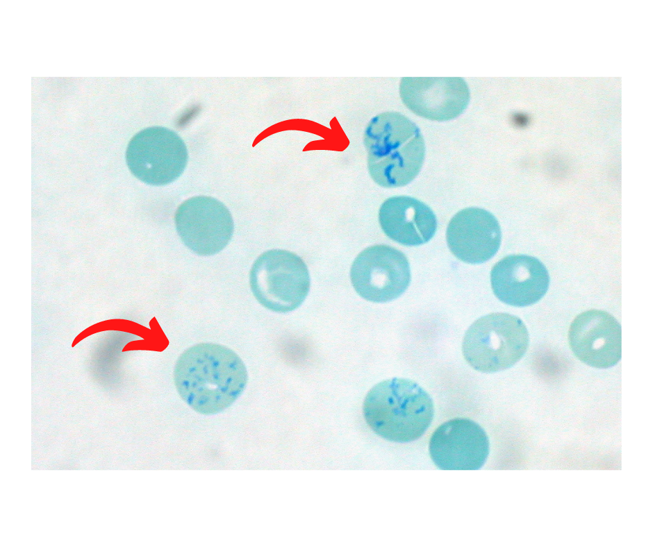
Image shows Supravital stain of a smear of human blood from a patient with hemolytic anemia. What is the cell marked by arrow?
 By Ed Uthman, MD, pathologist, Houston, Texas, USA - Own work, CC BY 3.0, https://commons.wikimedia.org/w/index.php?curid=12209264
Supravital stain of a smear of human blood :
From a patient with hemolytic anemia.
The reticulocytes are the cells with the dark blue dots and curved linear structures (reticulum) in the cytoplasm.
Hereditary spherocytosis is most commonly due to mutations in genes that code for -
spectrin (alpha and beta), ankyrin, band 3 protein, protein 4.2
Hereditary Spherocytosis autosomal recessive inheritance usually account for -
Recessive inheritance may account for 20-25% of all HS cases
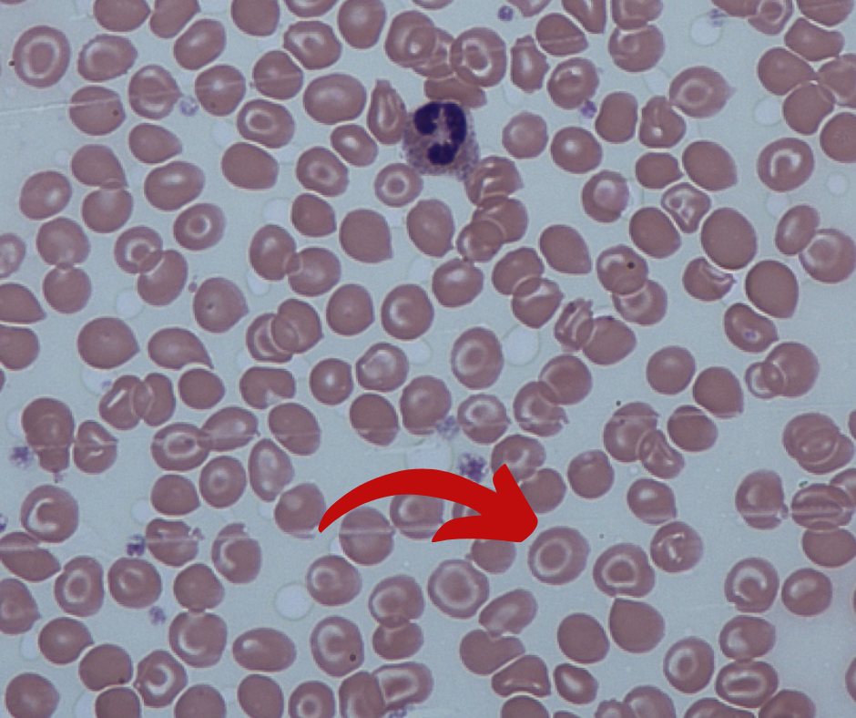
Which is the condition where cell marked in the IMAGE is seen most commonly ?
 By Dr Graham Beards - Own work, CC BY-SA 3.0, https://commons.wikimedia.org/w/index.php?curid=20526635
Target cells (codocytes): Most commonly seen in thalassemia.
Normal Hb has two alpha, and two beta chains, a decrease in alpha chains is alpha thalassemia, a decrease in beta chains is beta-thalassemia.
Target cells appear in association with the following conditions: Liver disease: Lecithin—cholesterol acyltransferase (LCAT) activity may be decreased in obstructive liver disease. Alpha-thalassemia and beta-thalassemia Hemoglobin C Disease Iron deficiency anemia Post-splenectomy Autosplenectomy
Amino acid substitution of lysine for glutamic acid at position six of the beta hemoglobin chain results in which of the following?
Hemoglobin S- coding of valine instead of glutamate in position 6 of the hemoglobin beta chain
Howell–Jolly body are seen in most commonly which of the following condition ?
Howell–Jolly body is a cytopathological finding of basophilic nuclear remnants (clusters of DNA) in circulating erythrocytes
Spleens are also removed for therapeutic purposes in conditions like hereditary spherocytosis, trauma to the spleen, and autosplenectomy caused by sickle cell anemia.
Myelodysplastic syndrome can transform into which of the following?
Myelodysplastic syndrome (MDS) often transforms into acute myeloid leukemia [AML].
Transformation of MDS into acute lymphoblastic leukemia (ALL) is extremely rare.
In Myelodysplastic syndrome if Bone-marrow myeloblasts rises over ---------% it is considered transformation to acute myelogenous leukemia [WHO] ?
Overall percentage of bone-marrow myeloblasts rises over a particular cutoff (20% for WHO and 30% for FAB), then transformation to acute myelogenous leukemia (AML) is said to have occurred.
Reticulocyte index for a healthy individual should be in range?
Reticulocyte index (RI) should be between 0.5% and 2.5% for a healthy individual.


