Image Question-6
Contents

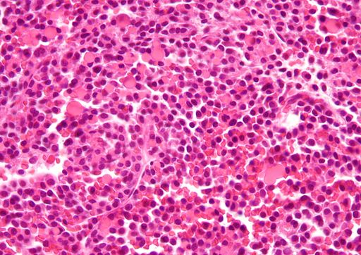
Russell bodies –

Above Image shows – Russell bodies
- Found in multiple myeloma.
- Russell bodies are inclusion bodies usually found in atypical plasma cells that become known as Mott cells.
- Russell body is characteristic of the distended endoplasmic reticulum.
Dutcher bodies
- Similar inclusion bodies like Russel bodies
- Overlie the nucleus or invaginate into Nucleus.
Extra-Points
Degmacyte or bite cell
- Bite cells are known to be a result from processes of oxidative hemolysis, such as Glucose-6-phosphate dehydrogenase deficiency,
- It is an abnormally shaped mature red blood cell with one or more semicircular portions removed from the cell margin, known as “bites”
- Macrophages in the spleen bite Heinz bodies out and the resulting red cells are actually called bite cells
Howell-Jolly bodies
- Howell-Jolly bodies – little fragments of the red cell nucleus.
- Most commonly seen in patients after splenectomy.
Heinz bodies
- Heinz bodies are seen in G6PD deficiency.
- Heinz bodies – represent denatured globin chains.


