Image Question-55
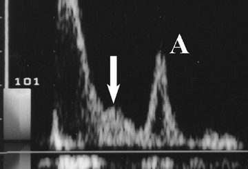
What is the most likely diagnosis from Echocardiogram?
https://drive.google.com/file/d/1v_RTwbM4Uy-lt173_vNlwYUz_TCSFNLy/view?usp=sharing
[A] RCM
[B] HOCM
[C] DCM
[D] ASD
Characteristic echocardiographic findings of RCM include:
- Biatrial enlargement: The atria are strikingly enlarged
- Diastolic dysfunction
- Normal or near-normal systolic function: The left ventricular (LV) and right ventricular ejection fraction are normal or mildly reduced
- Hypertrophy: The ventricles are hypertrophied with decreased compliance
- Mass-like apical lesions: These lesions are associated with restriction of LV and RV filling
- Mitral and tricuspid valve leaflet tethering: This may result in regurgitation
- Nondilated ventricles
- Doppler imaging shows a restrictive filling pattern with tissue Doppler showing an elevated E/e’ ratio.
Whatsapp Link
https://whatsapp.com/channel/0029VaA8rdB4Y9lmzS8S9Z2Q/1894
https://whatsapp.com/channel/0029VaA8rdB4Y9lmzS8S9Z2Q


