Image Question-5
What is the diagnosis of IMAGE?
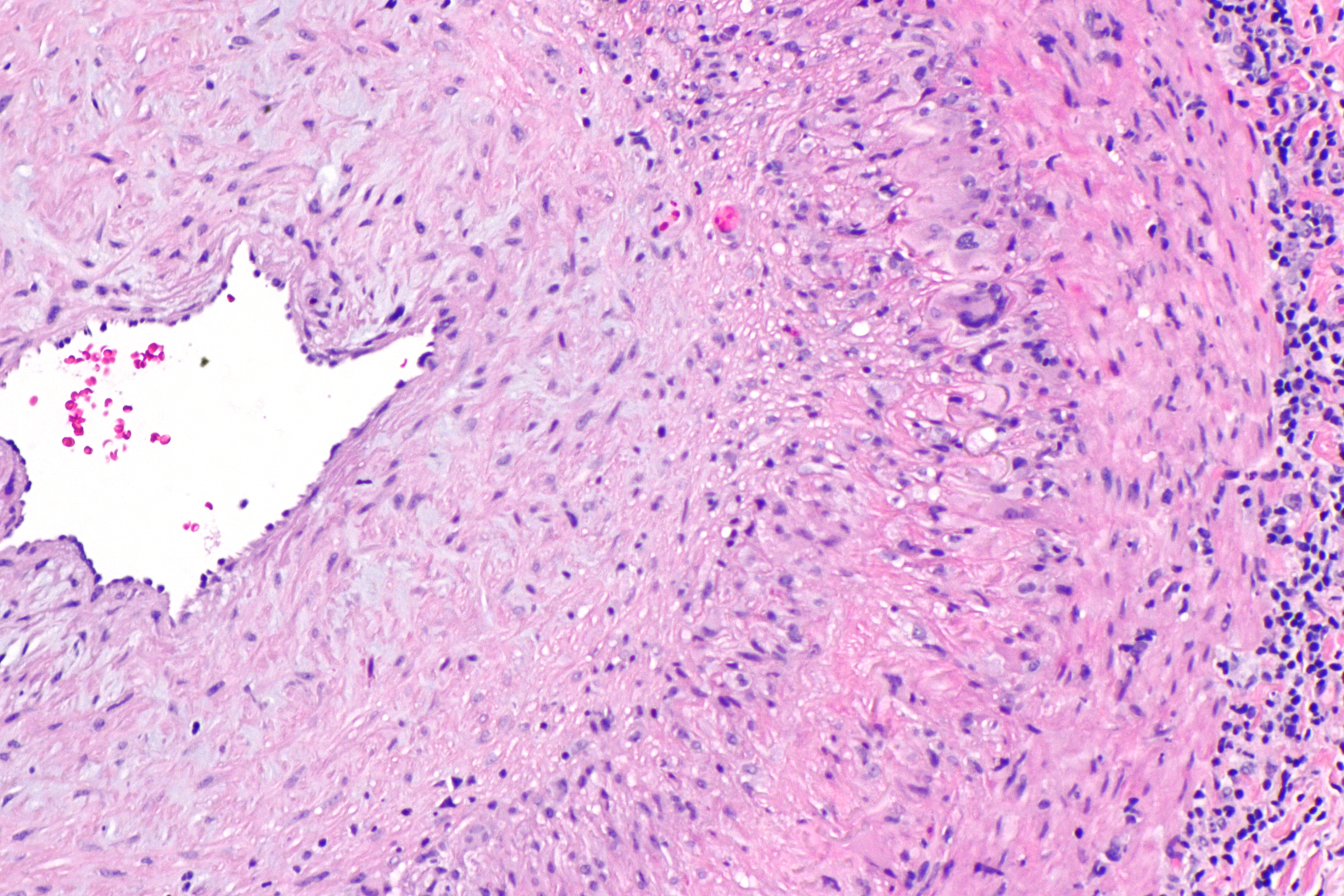
A 72 year female patient presented with frequent, severe headaches and tenderness over the temples. jaw pain while eating or talking. He also complained of double vision of both eyes. What is the most probable diagnosis?
A. SLE
B. Takayasu arteritis
C. Giant cell arteritis
D. Aortoarteritis
A. SLE
B. Takayasu arteritis
C. Giant cell arteritis
D. Aortoarteritis

IMAGE –
– The arterial lumen is seen on the left.
– A giant cell is seen on the right at the interface between the thickened intima and media.


