ECG Question-2
Contents
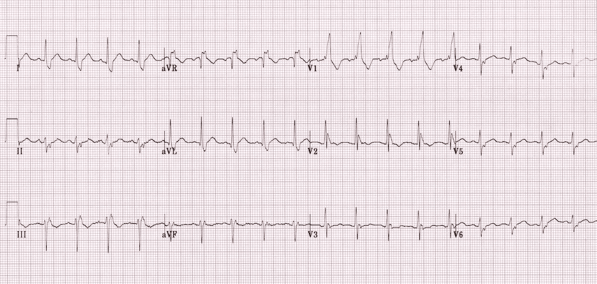
What is the diagnosis of ECG?
A. LBBB + LAFB
B. LBBB + LPFB
C. RBBB + LAFB
D. RBBB + LPFB
What is Bifascicular Block?
RBBB + LAFB is called Bifascicular Block ?
What are the types of bifascicular block [ECG patterns]?
- Right bundle branch block (RBBB) with left anterior fascicular block (LAFB) – left axis deviation (LAD)
- RBBB and left posterior fascicular block (LPFB) – right axis deviation (RAD) in the absence of other causes
| Bifascicular Block | ||
| 1 | RBBB + LAFB | More common |
| 2 | RBBB + LPFB | Less common |
Why RBBB + LAFB is more common than RBBB + LPFB in bifascicular block?
| Why commonest type of bifascicular block is RBBB + LAFB ? [ECG patterns] | Vulnerability to Damage |
| The LAF is supplied by a single branch of the left anterior descending artery (LAD). The LPF, on the other hand, receives a dual blood supply from both the right and left circumflex arteries. | Because the LAF has a single blood supply, it’s more susceptible to damage and block due to ischemia or other factors affecting the LAD. The LPF’s dual blood supply makes it more resistant to damage and block compared to the LAF. |
Anatomy and Blood Supply:
The LAF is supplied by a single branch of the left anterior descending artery (LAD).
The LPF, on the other hand, receives a dual blood supply from both the right and left circumflex arteries.
Vulnerability to Damage:
Because the LAF has a single blood supply, it’s more susceptible to damage and block due to ischemia or other factors affecting the LAD.
The LPF’s dual blood supply makes it more resistant to damage and block compared to the LAF.
Location and Relation to LV Outflow Tract:
The LAF’s location and relationship to the left ventricular (LV) outflow tract can also contribute to its vulnerability to damage, as it can be subjected to mechanical trauma.
Clinical Significance:
RBBB + LAFB is a common finding in patients with coronary artery disease (CAD), and its presence can indicate underlying structural and electrical abnormalities.
RBBB + LPFB is less common and may be associated with more extensive underlying cardiac pathology.
| Blood supply | |||
| Left anterior fascicle (LAF) | More common for block | Single blood supply | LAD |
| Left posterior fascicle (LPF) | Less common for block | Dual blood supply | RCA + LCX |
Arterial switch operation
Contents1 Which of the following is the Arterial switch operation?2 Jatene procedure is ideally performed...
Artery of Percheron
Contents1 Artery of Percheron is a branch of2 Artery of Percheron arises from which part...
Artery of Salmon
Contents1 Artery of Salmon is a branch of2 Artery of Salmon arises from which segment...
Artery of Wollschlaeger and Wollschlaeger
Which of the following is the ‘Artery of Wollschlaeger and Wollschlaeger’ [A] Occipital artery[B] Single...
Aschoff bodies
Aschoff bodies are nodules found in the hearts of individuals with rheumatic fever. Characteristic of...
ASD Flow across the defect
In ASD Flow across the defect occurs in [A] Mostly in Systole[B] Only diastole[C] Only...
ASD Flow across the defect in restrictive defect
ASD Flow across the defect in restrictive defect is determined largely by [A] Size of...
Atopic Dermatitis
Contents1 Skin in atopic dermatitis is deficient in2 The infectious agent most often involved in...
Atopic Dermatitis – Hanifin and Rajka criteria
Contents1 Atopic Dermatitis2 Hanifin and Rajka criteria3 Major criteria (need 3 or more) are as...