Dynamic outflow obstruction in HOCM
Contents
- 1 Dynamic outflow obstruction in HOCM is due to
- 2 Asymmetric left ventricular hypertrophy most commonly affects –
- 3 Most common complain in symptomatic cases in HOCM –
- 4 In Asymmetric septal hypertrophy septal thickness is ————- the thickness of the posterior wall
- 5 Banana-shaped LV cavity seen in –
- 6 Systolic anterior motion
- 7 Mechanism of SAM
- 8 Echocardiographic features – In most common forms of HCM of the obstructive type-
Dynamic outflow obstruction in HOCM is due to
A. Infundibular hypertrophy
B. Systolic anterior motion
C. Subaortic membrane
D. High Blood flow through LVOT
Asymmetric left ventricular hypertrophy most commonly affects –
A. RV
B. LV Free wall
C. Ventricular septum
D. LV Apex
Most common complain in symptomatic cases in HOCM –
A. Chest pain
B. Palpitation
C. Syncope
D. Dyspnea
In Asymmetric septal hypertrophy septal thickness is ————- the thickness of the posterior wall
A. ≥ 0.9 times
B. ≥ 1.5 times
C. ≥ 1.6 times
D. ≥ 1.8 times
Banana-shaped LV cavity seen in –
A. HCM
B. RCM
C. DCM
D. Aortic stenosis
Systolic anterior motion
- Dynamic outflow obstruction in HOCM is due to systolic anterior motion(SAM) of the anterior leaflet of the mitral valve.
- This is due to impingement of the mitral valve leaflets on the hypertrophied basal septum.
- The outflow tract obstruction is dynamic and caused by a pressure gradient which pulls the anterior leaflet of the mitral valve anteriorly further leading to outflow tract obstruction.
- It can occur in patients with and without hypertrophic cardiomyopathy.
Mechanism of SAM
Mechanisms for SAM
- Venturi effect
- Drag effect
Both mechanisms describe the anterior leaflet being drawn into the outflow tract either by a pulling (venturi) or pushing (drag) phenomenon
Echocardiographic features – In most common forms of HCM of the obstructive type-
- Small, hyperdynamic left ventricle
- Thick sigmoid septum
- Banana-shaped cavity,
- Asymmetric septal hypertrophy (septal thickness ≥1.6 times the thickness of the posterior wall),
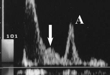
- Relatively small LVOT, elevated flow velocity in the LVOT that peaks in late systole
(when the LVOT is smallest), - Systolic anterior motion of the mitral valve,
- Significant amount of posteriorly directed MR


