Dot-Dash sign
Contents
- 1 Which of the following condition is associated with ‘Dot-Dash sign’?
- 2 Which is the primary technique for identifying white matter lesions caused by multiple sclerosis?
- 3 ‘Dot-Dash’ sign
- 4 ‘Dot-Dash’ sign
- 5 Clinical significance –
- 6 Most sensitive single finding for multiple sclerosis in the MR imaging-
- 7 What is the histopathologic basis of the Dot-Dash sign presence in early multiple sclerosis?
Which of the following condition is associated with ‘Dot-Dash sign’?
A. Motor Neuron Disease
B. Multiple sclerosis
C. Alzheimer’s disease
D. Spinal Muscular Atrophy
Which is the primary technique for identifying white matter lesions caused by multiple sclerosis?
A. CT Scan
B. PET-CT
C. Cerebral Angiography
D. MR imaging
‘Dot-Dash’ sign
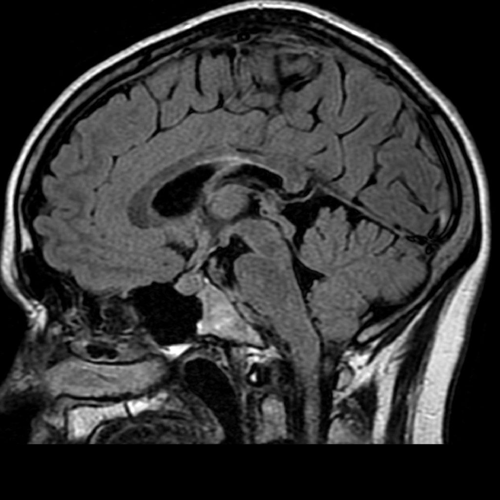
MR imaging finding that is also highly sensitive for multiple sclerosis—an ependymal irregularity -“Dot-Dash” sign.
This sign appears to suggest a diagnosis of multiple sclerosis at an earlier stage
‘Dot-Dash’ sign correlates with ependymal perivenular inflammation-an early histopathological event in multiple sclerosis-which is a more sensitive biomarker for the early diagnosis of multiple sclerosis via MRI technology.
‘Dot-Dash’ sign
Look at the – High signal along the ependymal surface

Case courtesy of Frank Gaillard, Radiopaedia.org. From the case rID: 9941
- Dot-Dash’ sign is best seen on sagittal FLAIR
- Seen along the inferior surface of the corpus callosum and roof of the lateral ventricle bodies.
- It is seen as tiny (~1 mm) dots of high signal along the ependymal surface that may coalesce into short dash.
Clinical significance –
Dot-Dash’ sign – distinguish multiple sclerosis from neuromyelitis optica which less frequently demonstrate this sign.
Presence of Dot-Dash’ sign suggest – Multiple sclerosis
Most sensitive single finding for multiple sclerosis in the MR imaging-
Subcallosal striations
What is the histopathologic basis of the Dot-Dash sign presence in early multiple sclerosis?
Early multiple sclerosis lesions start around the subependymal venous wall


