Medicine Review MCQs -XIII
Contents
- 1 All of the following are CORRECT for Bernard–Soulier syndrome EXCEPT -
- 2 What is the Marked structure in Image?
- 3 Platelet-producing megakaryocytes go through ................during cell differentiation
- 4 Megakaryocytes are derived from ................... stem cell
- 5 The primary signal for megakaryocyte production is -
- 6 All of the following include Lymphoid cells EXCEPT -
- 7 Clock Face Nucleus seen in -
- 8 Reed-Sternberg cells seen in -
- 9 Owl’s Eye Appearance nucleus seen in -
- 10 Owl's eye appearance of the Lentiform nucleus of the basal ganglia seen on head CT scan images of patients with -
All of the following are CORRECT for Bernard–Soulier syndrome EXCEPT -
Bernard–Soulier syndrome
Low platelet count
Bernard–Soulier syndrome -caused by a deficiency of the glycoprotein Ib-IX-V complex (GPIb-IX-V), the receptor for von Willebrand factor
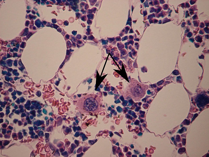
What is the Marked structure in Image?
 By WVSOM_Megakaryocytes.JPG: Wbensmithderivative work: Icewalker cs (talk) - WVSOM_Megakaryocytes.JPG, CC BY 3.0, https://commons.wikimedia.org/w/index.php?curid=15216382 - Medicine Question Bank
Megakaryocytes in bone marrow -marked with arrows
Platelet-producing megakaryocytes go through ................during cell differentiation
Platelet-producing megakaryocytes go through endomitosis during cell differentiation
Megakaryocytes are derived from ................... stem cell
Megakaryocytes are derived from hematopoietic stem cell precursor cells in the bone marrow. They are produced primarily by the liver, kidney, spleen, and bone marrow.
The primary signal for megakaryocyte production is -
The primary signal for megakaryocyte production is thrombopoietin
sufficient but not absolutely necessary for inducing differentiation of progenitor cells in the bone marrow towards a final megakaryocyte phenotype.
All of the following include Lymphoid cells EXCEPT -
Lymphoid cells include T cells, B cells, natural killer cells, and innate lymphoid cells.
Clock Face Nucleus seen in -
Plasma cell nucleus - cartwheel or clock face arrangement
Reed-Sternberg cells seen in -
Hodgkin lymphoma characteristically presents with Hodgkin and Reed-Sternberg cells.
When the cells are mononucleated, they are called Hodgkin cells.
When they are multinucleated, they are called Reed-Sternberg cells.
Owl’s Eye Appearance nucleus seen in -
Owl's eye appearance of the Lentiform nucleus of the basal ganglia seen on head CT scan images of patients with -
Cerebral hypoxia


